Carregar apresentação
A apresentação está carregando. Por favor, espere

1
Hospital Sírio Libanês
GERME – Ago/ 2015 Hospital Sírio Libanês Eduardo Luis Bizetto Marcelo Bordalo Rodrigues Conrado Furtado Cavalcanti Denise Tokechi Amaral Rodrigo Yacubian Fernandes Marcos Felippe de P. Correa Ceci Obara Kurimori Hugo Pereira Costa Paulo Victor P. Helito Guilherme Echeverria Nasser

2
Caso 01 Fem, 34 anos dor na região plantar do II e III raios há 03 semanas. Nega trauma.

7
Caso 02 Fem, 34 anos dor na região plantar do 2º metatarso há 01 ano.
Nega trauma.

11
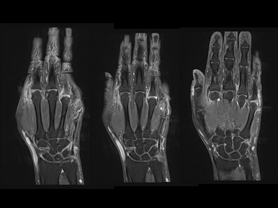
Caso 03 Masc, 58 anos dor e edema nas mãos e punhos há 08 meses.

15
Dactilites Artrites Psoriásicas

16
Dactilites Ocorre em cerca de 40% dos casos de artrite psoriásica;
Achado principal tenossivite dos flexores; Estudos baseados em RM demonstraram associação com entesites, edema periarticular e sinovite.

17
Dactilites Ocorre em cerca de 40% dos casos de artrite psoriásica;
Achado principal tenossivite dos flexores; Estudos baseados em RM demonstraram associação com entesites, edema periarticular e sinovite.

19
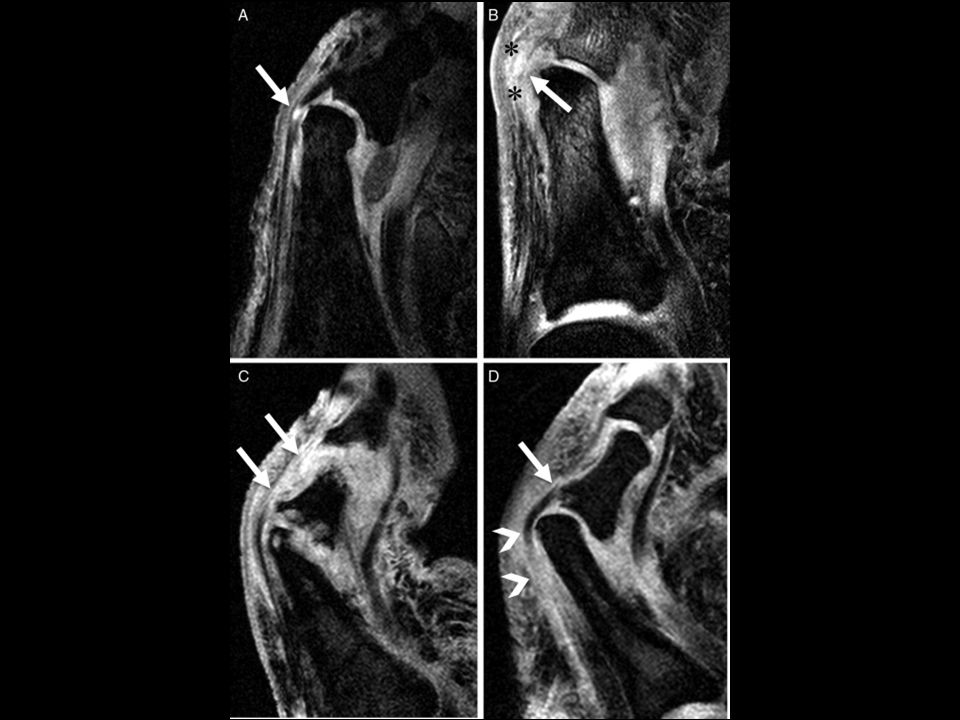
Alterações nas enteses
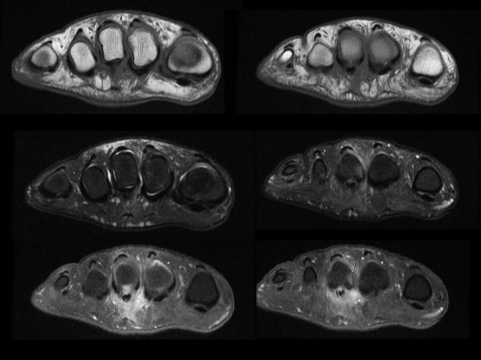
Dactilites Alterações nas enteses Inserção dos ligamentos colaterais 75% Inserção do tendão extensor 50% Realce da placa plantar 40% Nenhum paciente apresentou sinais de entesite na inserção do tendão flexor.

20
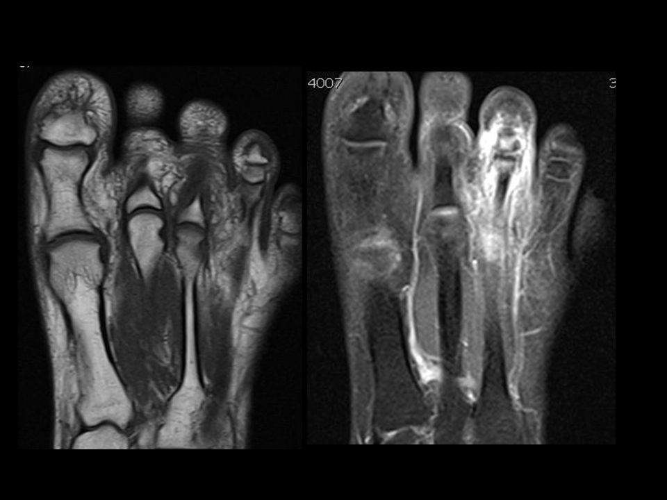
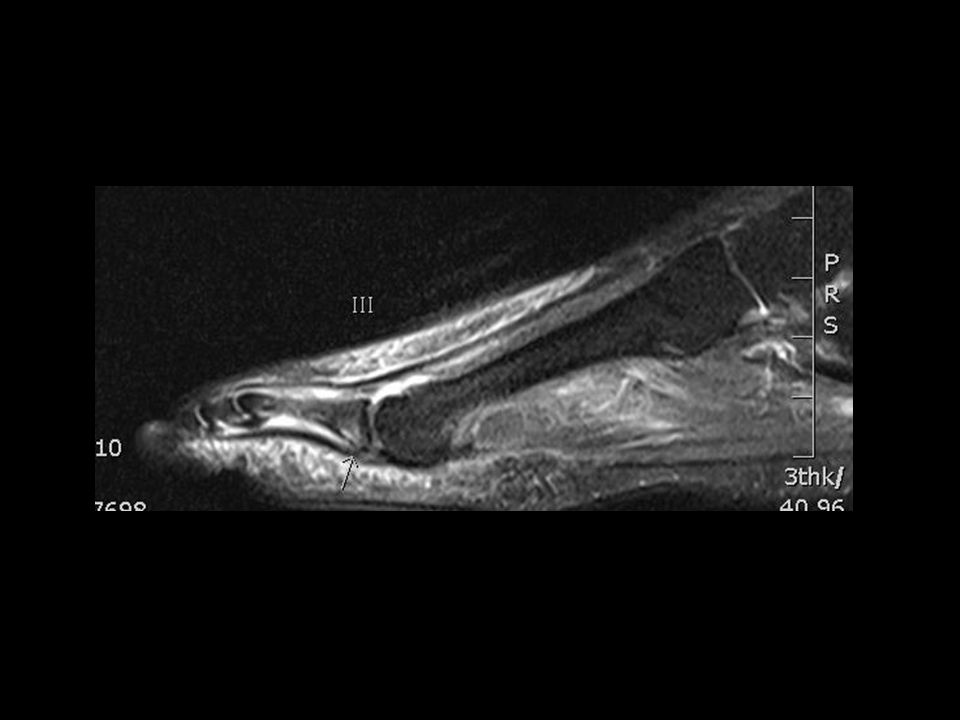
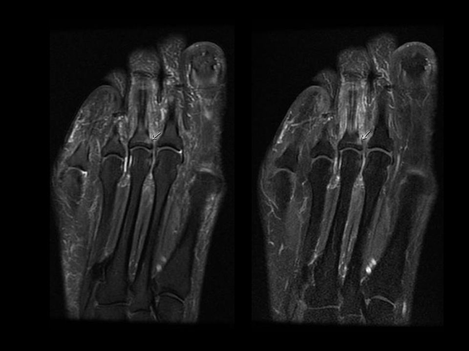
1 Enthesitis and osteitis in dactylitis
1 Enthesitis and osteitis in dactylitis. Fat-saturated contrast-enhanced T1-weighted images. (A) Coronal image of the left big toe of a 37-year-old man. Bilateral collateral ligament insertions and origins show mild to moderate enhancement (arrows). Mild to moderate bone marrow enhancement adjacent to the origins and insertions of the ligaments can also be seen (asterisks). (B) Sagittal image of the left big toe of same patient as (A) showing focal bone marrow enhancement at the extensor tendon insertion (arrow) and diffuse enhanced bone marrow (asterisk). Intracapsular synovium of the interphalangeal joint was swollen and moderately enhanced (arrowheads). (C) Sagittal image of the right middle finger of a 41-year-old man. The extensor tendon insertion demonstrated mild enhancement (arrow) with associated bone oedema (white outlined arrowhead). Diffuse enhancement of the flexor tenosynovium (white asterisks), extracapsular soft tissues (black asterisk), and moderate enhanced intracapsular synovium (arrowhead) in the proximal interphalangeal joint were also seen. (D) Coronal image of the right 2nd toe of a 42-year-old woman. The base of the middle phalangeal bone was partially enhanced (arrow), adjacent to the extensor tendon insertion. Diffuse soft tissue enhancement was also shown (black asterisks).
 Coronal image of the left big toe of a 37-year-old man. Bilateral collateral ligament insertions and origins show mild to moderate enhancement (arrows). Mild to moderate bone marrow enhancement adjacent to the origins and insertions of the ligaments can also be seen (asterisks). (B) Sagittal image of the left big toe of same patient as (A) showing focal bone marrow enhancement at the extensor tendon insertion (arrow) and diffuse enhanced bone marrow (asterisk). Intracapsular synovium of the interphalangeal joint was swollen and moderately enhanced (arrowheads). (C) Sagittal image of the right middle finger of a 41-year-old man. The extensor tendon insertion demonstrated mild enhancement (arrow) with associated bone oedema (white outlined arrowhead). Diffuse enhancement of the flexor tenosynovium (white asterisks), extracapsular soft tissues (black asterisk), and moderate enhanced intracapsular synovium (arrowhead) in the proximal interphalangeal joint were also seen. (D) Coronal image of the right 2nd toe of a 42-year-old woman. The base of the middle phalangeal bone was partially enhanced (arrow), adjacent to the extensor tendon insertion. Diffuse soft tissue enhancement was also shown (black asterisks).")
21
Alterações extracapsulares e sinovais
Dactilites Alterações extracapsulares e sinovais Edema pericapsular difuso 92% Sinais de sinovite 68% Aumento de sinal tendão extensor 42%

23
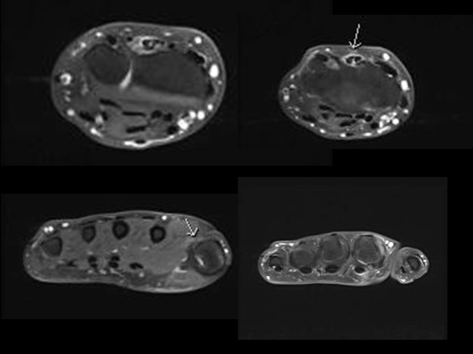
Alterações nos tendões flexores e polias anulares
Dactilites Alterações nos tendões flexores e polias anulares Tenossinovite dos flexores 75% Espessamento e alteração de sinas das polias 50%

24
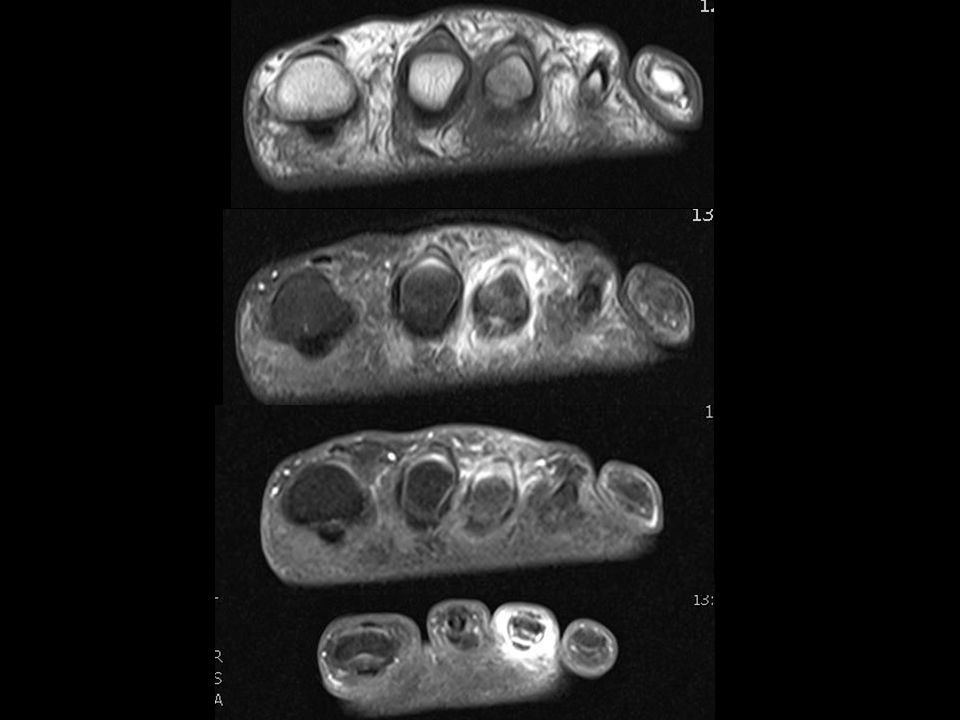
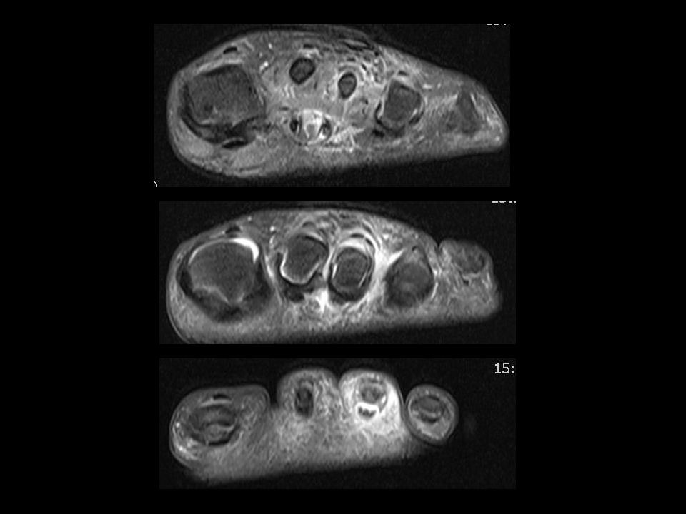
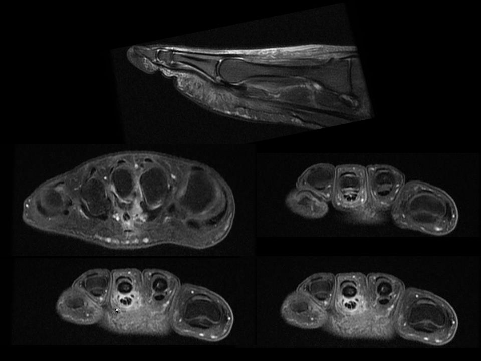
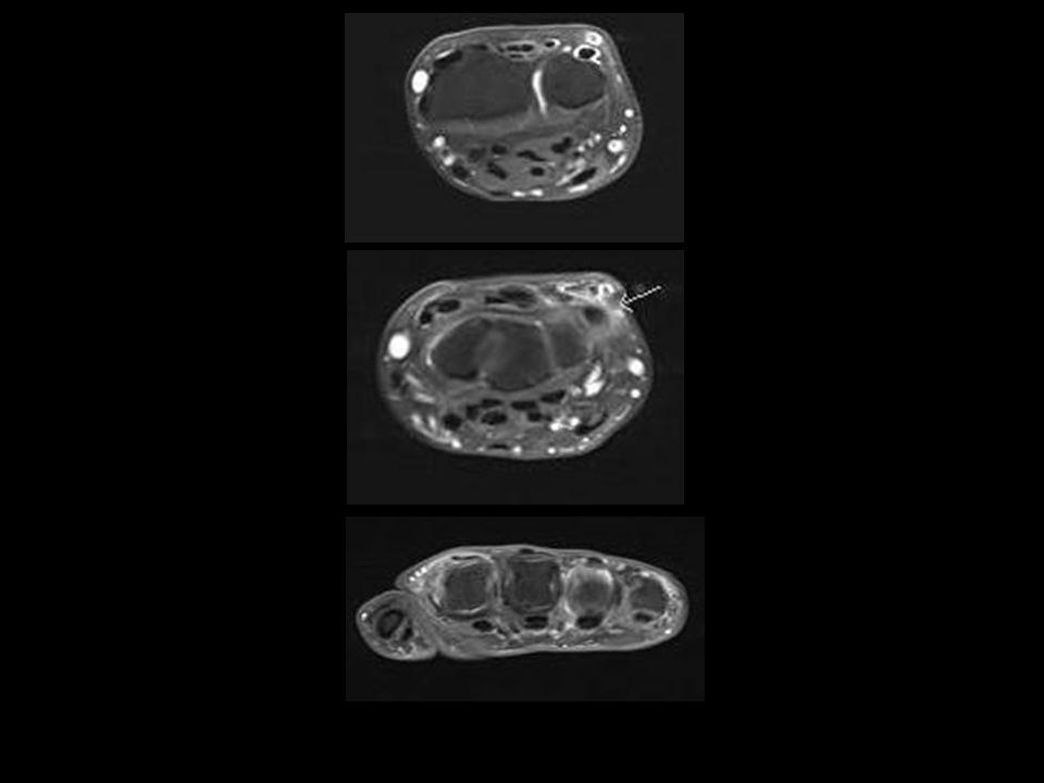
A2 A4 3 Finger pulleys and toe fibrous sheath abnormalities. Fat-saturated contrast-enhanced T1-weighted axial images. (A) Interphalangeal joint of the left thumb of a 57-year-old man. The A2 pulley region adjacent to the bone was unilaterally oedematous and ill-defined (white arrow). Area of enhanced high signal representing thickened flexor tenosynovium is also noted (arrowhead). (B) Distal site of proximal interphalangeal (PIP) joint of the left index finger of a 62-year-old man. There is oedematous change at bilateral A4 pulley attachments (white arrows). (C) PIP joint of the left index finger of same patient as (B). Symmetrical rim-like enhancement is observed around the A3 pulley (white arrows). The flexor tenosynovium is thickened as noted by the area of high signal (arrowhead). (D) PIP joint of the right 2nd toe of same patient as (2C) showing oedematous change of the fibrous sheath (white arrows). A3 Bainha
 Interphalangeal joint of the left thumb of a 57-year-old man. The A2 pulley region adjacent to the bone was unilaterally oedematous and ill-defined (white arrow). Area of enhanced high signal representing thickened flexor tenosynovium is also noted (arrowhead). (B) Distal site of proximal interphalangeal (PIP) joint of the left index finger of a 62-year-old man. There is oedematous change at bilateral A4 pulley attachments (white arrows). (C) PIP joint of the left index finger of same patient as (B). Symmetrical rim-like enhancement is observed around the A3 pulley (white arrows). The flexor tenosynovium is thickened as noted by the area of high. signal (arrowhead). (D) PIP joint of the right 2nd toe of same patient as (2C) showing oedematous change of the fibrous sheath (white arrows). A3. Bainha.")
25
Dactilites Conclusão:
As entesites comuns na artrite psoriásica também tem papel importante nas dactilites. Entesopatia funcional das polias dos flexores e bainha fibrosa poderia explicar a tenossinovite dos flexores Não se observa entesite dos flexores Alterações de ênteses funcionais poderiam explicar as alterações inflamatórias extracapsulares

26
Referências Bibliográficas
Tan AL, Fukuba E, Halliday NA, Tanner SF, Emery P, McGonagle D. High-resolution MRI assessment of dactylitis in psoriatic arthritis shows flexor tendon pulley and sheath-related enthesitis. Ann Rheum Dis Jan;74(1):185-9. Ritchlin CT. Psoriatic enthesitis: an update from the GRAPPA 2013 Annual Meeting. J Rheumatol 2014;41:1220–3.
: Ritchlin CT. Psoriatic enthesitis: an update from the GRAPPA 2013 Annual Meeting. J Rheumatol 2014;41:1220–3.")
Apresentações semelhantes


>")


