Carregar apresentação
A apresentação está carregando. Por favor, espere

1
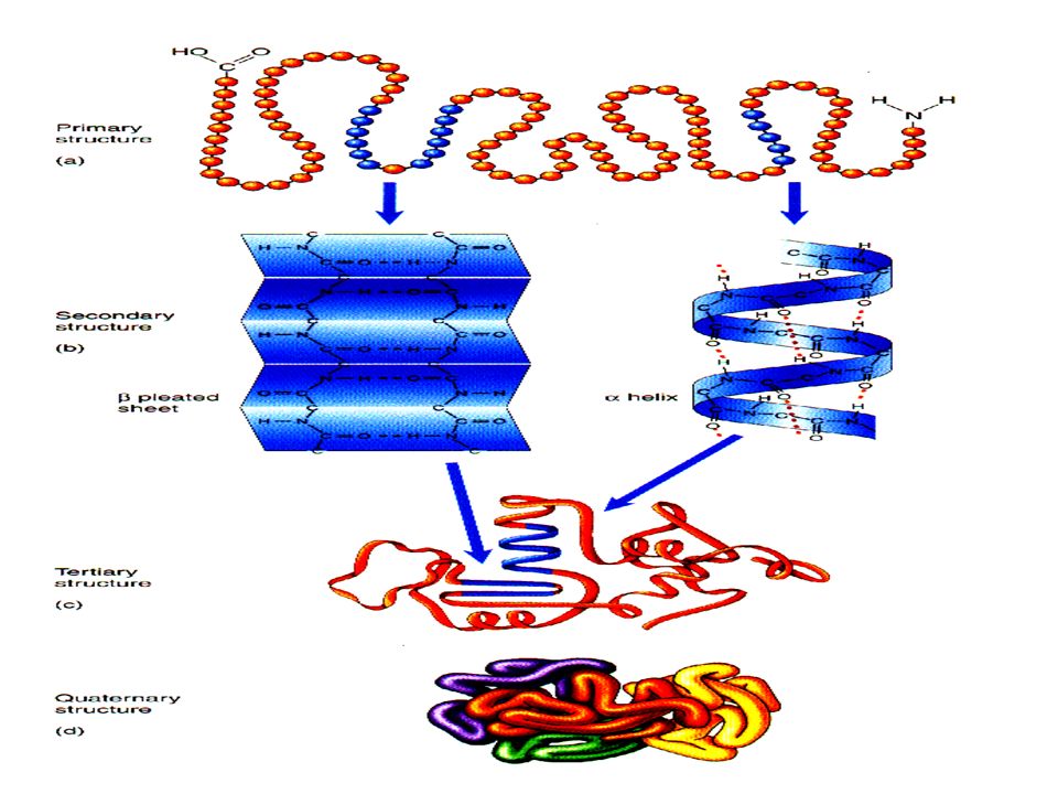
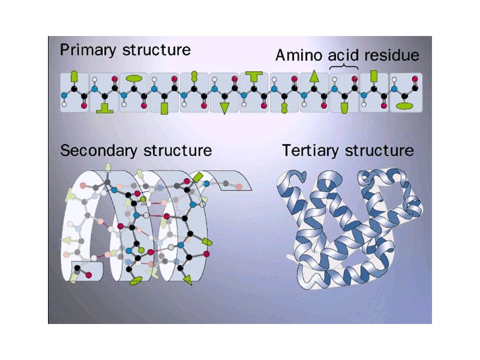
Estrutura de Proteínas

4
Figure 2.5. O pI é o pH onde a carga líquida da proteína é ZERO. A pH’s menores a proteína tem carater CATIÔNICO e a pH’s acima do pI aniônico. Lembra que pKa é “invariável” o termo variável é o pH.

5
Força Iônica e Enovelamento de Proteínas. AA´s básico e ácidos

7
-S-S-, H///O, cargas, VDW, uréia A partir da seq. 1ária, possibilidades de enovelamento enorme!! Como sabe?? Proteínas que ajudam no Enovelamento “Folding”

8
Efeito de íons, Espcificidade de íons, Caotrópicos e Cosmotrópicos Denaturam “Naturam”

13
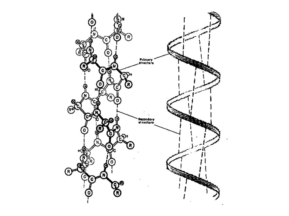
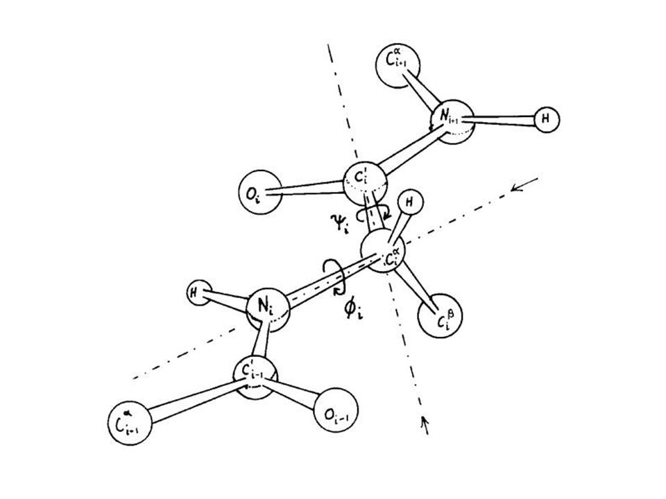
1 = 1,5 Å Anstieg pro Aminosäurerest 2 = 5,4 Å = 3,6 Aminosäurereste pro Windung 3 = 27 Å entspricht 18 Aminosäurereste auf 5 Windungen 4 = 26° 5 = 5,1 Å w1 - w5 = je 1 Helix-windung

14
3,6 resíduos de aminoácido por “passo” ~ 5.4 ª
Estabilizado por H-ponte entre. Como devem ser os grupos R? e o interior da Hélice??

17
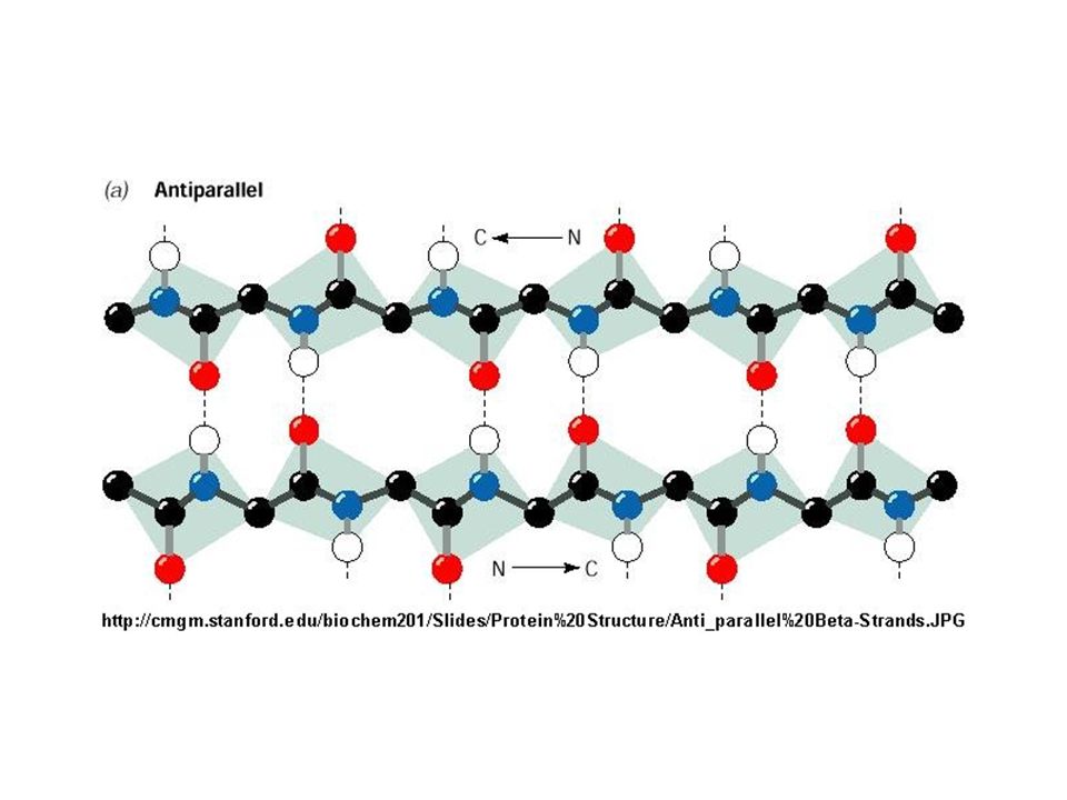
. Folha anti paralela

26
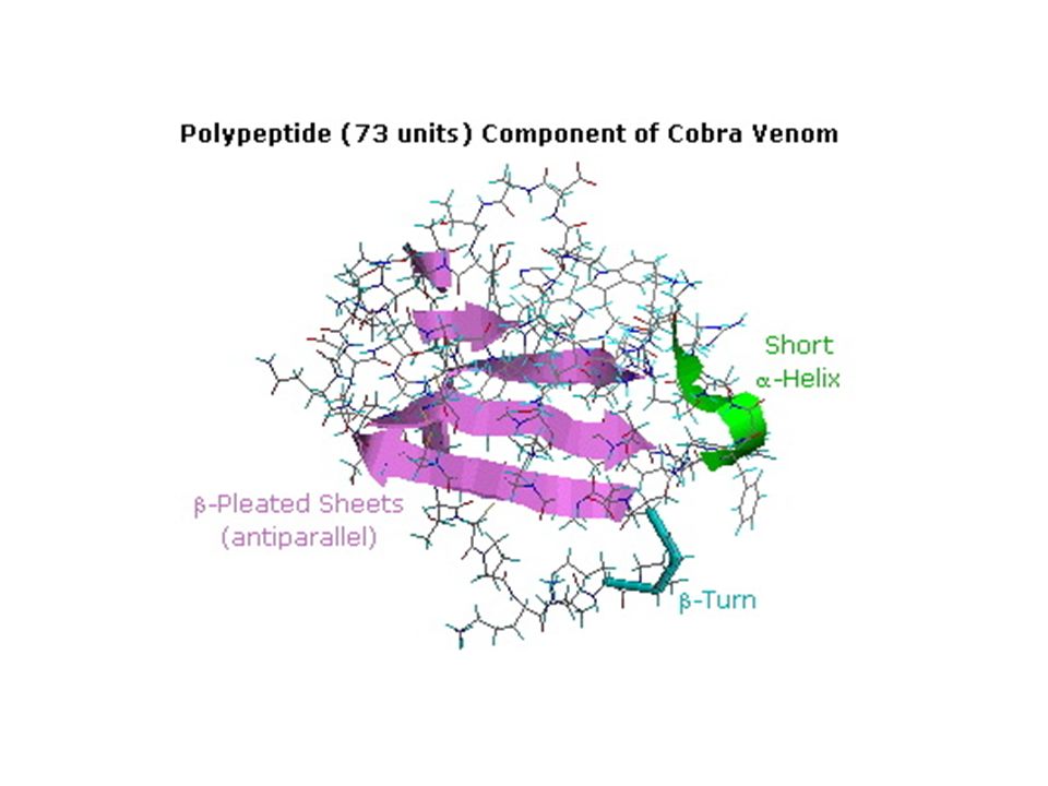
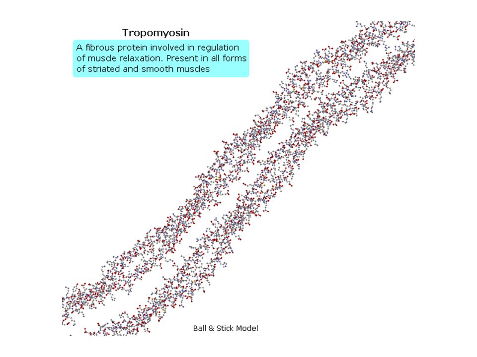
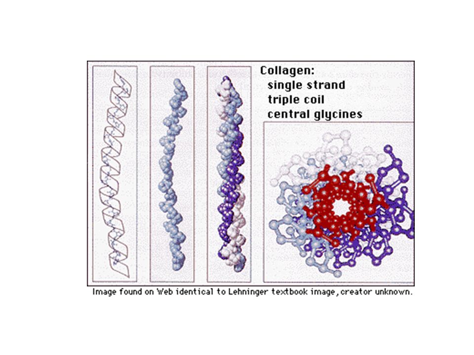
endothelin angiogenin lysozyme mellitin collagen thioredoxin hemoglobin

30
Brookhaven Protein Data Bank
The Brookhaven Data Bank will contain the most recent entries of all protein structural data. Proteins can easily be searched by using Brookhaven's PDB Gopher. When the box appears type in any key word. If a match appears, select the protein of your choice. Three files will appear: *.biblio, *.full, and *.gif. The first file will only give the information found in the header, it will not give coordinate data, the *.full file gives all data and is needed if you wish to manipulate structures from your own terminal. The *.gif file will contain a single image of the molecule. Remember that many cases several structures will be found for the same keyword. As an example try typing in pepsin after clicking here Next click on one of the many structures that show up on screen. Take a look at each of the files (*.biblio, *.full, *.gif) that are available from Brookhaven. For more information about the Brookhaven Protein Data Base connect to their home directory which contains FTP access to recent PDB Newsletter. A searchable index by key words can be used to search the Newsletter.
 that are available from Brookhaven. For more information about the Brookhaven Protein Data Base connect to their home directory which contains FTP access to recent PDB Newsletter. A searchable index by key words can be used to search the Newsletter.")
31
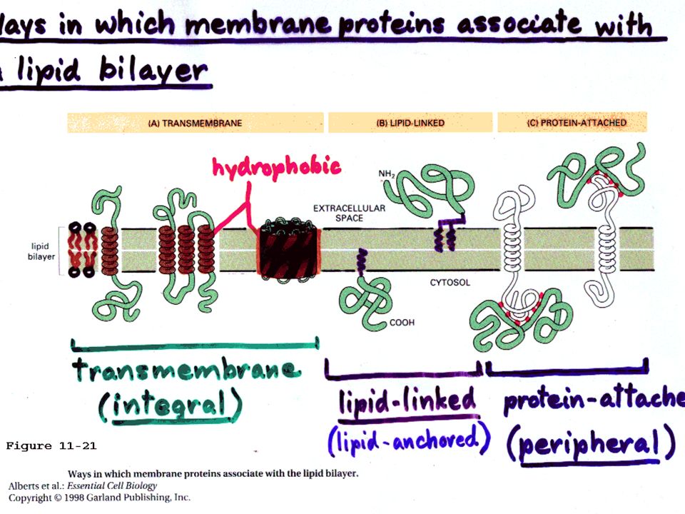
Proteínas de membrana:
-Integrais -Periféricas

35
Figure 4.19

44
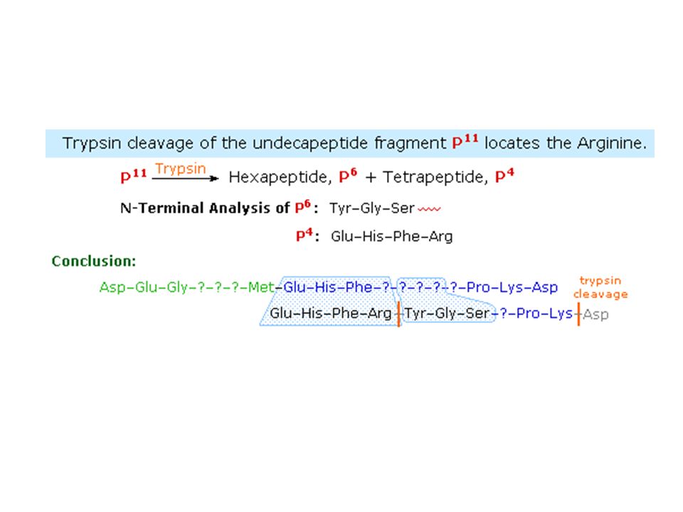
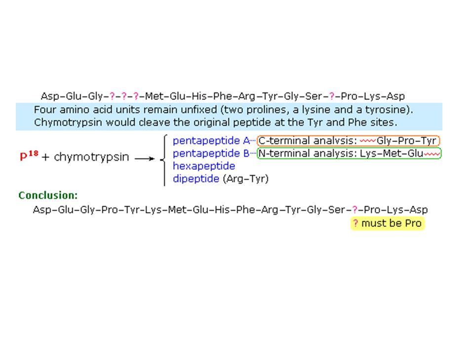
Carboxyl Side of Methionine
Name Type Specificity Cyanogen Bromide Chemical Carboxyl Side of Methionine Trypsin Enzymatic Carboxyl Side of Basic Amino Acids e.g. Lys & Arg Chymotrypsin Carboxyl Side of Aryl Amino Acids e.g. Phe, Tyr & Trp

50
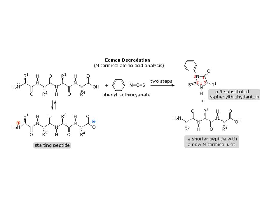
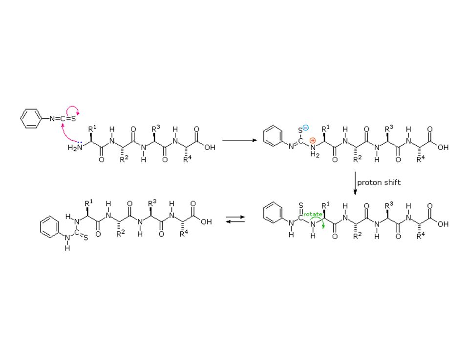
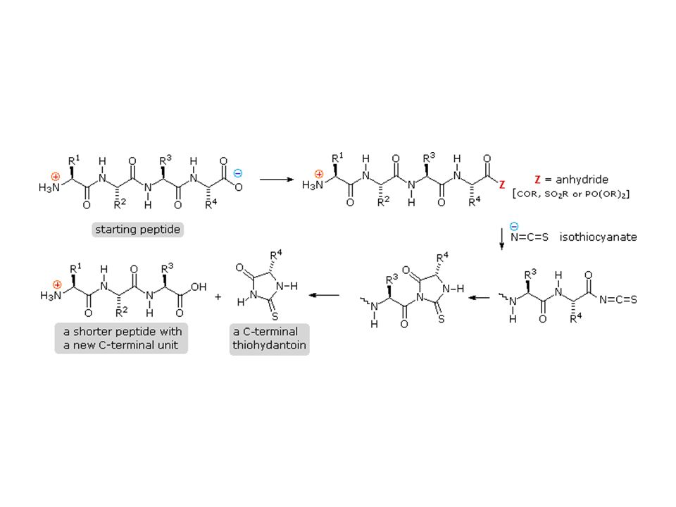
Síntese de Peptídeos

52
The Merrifield Peptide Synthesis

53
The Merrifield Peptide Synthesis

56
Copyright 2002 - 2007 thermus.org. All rights reserved
Copyright thermus.org. All rights reserved

61
HCO3- + H+ shifts --->to H2CO3 s & vice versa
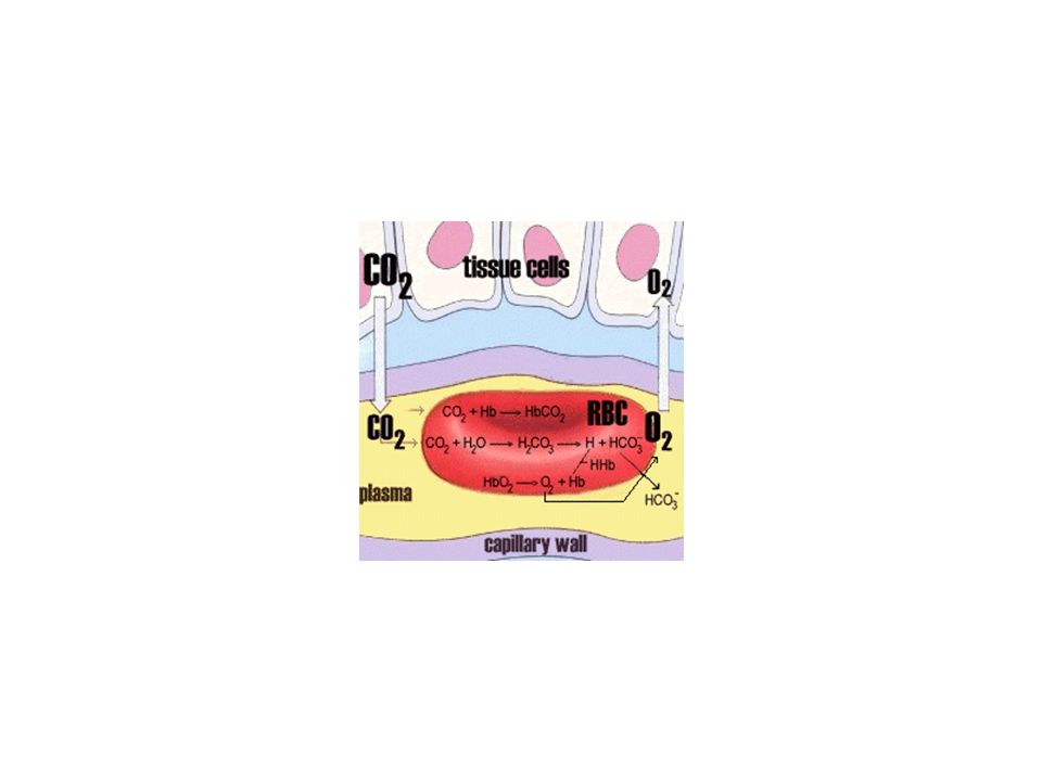
pH regulação do pH do sangue pH 7.4 + 0.1 Uma mudança de 0.4 unidades = “Morte” !!! Síndrome de Andrômeda- micróbio virulento do espaço infecta pessoas de cidade interiorana dos E.U.A. Todos morrem de “coagulação” do sangue !!! Curva de crescimento do micróbio = faixa estreita de pH!!! Somente dois sobreviventes - Um bebe que não parava de chorar, e assim exalava CO2 alkalose - Um bêbedo que tinha ulcera gastrica aberta = acidose. Anidrase Carbônica CO2 + H2O <--AC--> H2CO3 <---> H+ + HCO3- Hb “pega” H+ ions... tamponando blood cell... Se pH cai [H+ ^] então Anidrase carbonica HCO3- + H+ shifts --->to H2CO3 s & vice versa

65
Imidazol

68
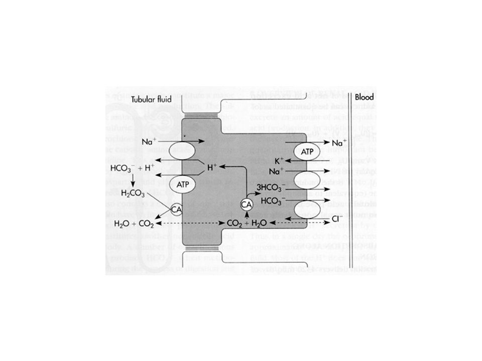
Informações: Fosfato tampão importante dentro das células e Rins Anidrase carbônica Localisada no citosol de células (renal e hemácias). Ao longo “the brush border” of the proximal tubule Rim. Não encontrada no plasma. pKa da Hb é funçao da ligação de O2 Em Fisiologia será estudado detalhes da liberação de H+ e outras Por rim, pulmão etc..

70
No metabolismo aeróbico cerca de 0.8 equivalentes de CO2
são produzidos por O2 consumido. Maior parte transportado como Bicarbonato! (anidrase carbônica) Restante carregado pela Hb como Carbamato
 Restante carregado pela Hb como Carbamato.")
Apresentações semelhantes


>")
