Carregar apresentação
A apresentação está carregando. Por favor, espere

1
ESTRUTURA GERAL DO TELENCÉFALO

2
CONTEÚDO 1- ASPECTOS GERAIS 2- OS LOBOS CEREBRAIS
3- OS NÚCLEOS DA BASE 4- O SISTEMA VENTRICULAR 5- ANATOMIA FUNCIONAL DO CÓRTEX CEREBRAL 6- O SISTEMA LÍMBICO 7- A CITOARQUITETURA DO CÓRTEX CEREBRAL

3
ASPECTOS GERAIS

4
RELEMBRANDO PROSENCÉFALO MESENCÉFALO ROMBENCÉFALO TELENCÉFALO
DIENCÉFALO MESENCÉFALO METENCÉFALO MIELENCÉFALO

5
TELENCÉFALO

6
PARTES CONSTITUINTES DO TELENCÉFALO:
LOBOS CEREBRAIS SISTEMA LÍMBICO NÚCLEOS DA BASE

7
OS LOBOS CEREBRAIS

8
LOBOS CEREBRAIS FRONTAL PARIETAL ÍNSULA TEMPORAL OCCIPITAL TELENCÉFALO

9
LOBOS CEREBRAIS

10
LOBOS CEREBRAIS LOBO FRONTAL LOBO PARIETAL LOBO OCCIPITAL
LOBO TEMPORAL LOBO LÍMBICO GIRO DO CÍNGULO GIRO PARAHIPOCAMPAL HIPOCAMPO

11
LOBOS CEREBRAIS GIRO DO CÍNGULO GIRO PARAHIPOCAMPAL HIPOCAMPO

12
LOBOS CEREBRAIS

13
PRINCIPAIS CISURAS CEREBRAIS
SULCO CENTRAL SULCO LATERAL

14
ANATOMIA FUNCIONAL DO CÓRTEX CEREBRAL

15
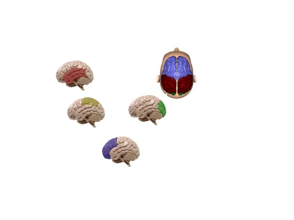
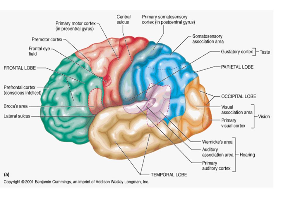
ÁREAS CEREBRAIS ESPECÍFICAS

16
OS NÚCLEOS DA BASE

17
OS NÚCLEOS DA BASE ESTRIADO: NÚCLEO CAUDADO - GLOBO PÁLIDO
NUCLEO LENTIFORME PUTAMEN

18
OS NÚCLEOS DA BASE

19
OS NÚCLEOS DA BASE

20
O SISTEMA VENTRICULAR

21
O SISTEMA VENTRICULAR VENTRÍCULOS LATERAIS AQUEDUTO CEREBRAL
TERCEIRO VENTRICULO QUARTO VENTRÍCULO

22
O SISTEMA LÍMBICO

23
O SISTEMA LÍMBICO -ÁREA SEPTAL -BULBO OLFATÓRIO -CORPO MAMILAR
-AMIGDALA -HIPOCAMPO -GIRO DO CÍNGULO

24
A CITOARQUITETURA DO CÓRTEX CEREBRAL

25
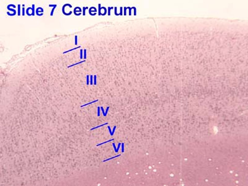
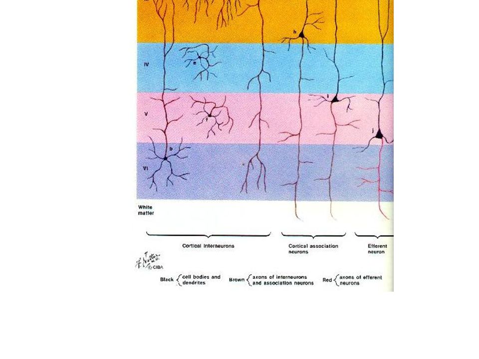
CITOARQUITETURA DO CÓRTEX CEREBRAL
I- CAMADA MOLECULAR II CAMADA GRANULAR EXTERNA III- CAMADA PIRAMIDAL EXTERNA IV- CAMADA GRANULAR INTERNA V-CAMADA PIRAMIDAL INTERNA VI- CAMADA CELULAS FUSIFORMES INTERNEURÔNIOS CORTICAIS NEURÔNIOS EFERENTES NEURÔNIOS DE ASSOCIAÇÃO CORTICAL

27
esquizofrenia INFECÇÕES VIRAIS MATERNAS NO QUINTO MÊS DE GESTAÇÃO
PODEM IFLUIR NA CORRETA MIGRAÇÃO DE NEURÔNIOS PA- RA A FORMAÇÃO DAS CAMADAS CORTICAIS E PREDISPOR A PES- SOA PARA A ESQUIZOFRENIA

28
I- CAMADA MOLECULAR POSSUI POUCAS CÉLULAS QUE SÃO CHAMADAS DE...
CÉLULAS HORIZONTAIS (OU DE CAJAL) QUE SÃO... NEURÔNIOS INTRACORTICAIS DE ASSOCIAÇÃO QUE POSSUEM... DENTRITOS E AXÔNIOS NA DIREÇÃO HORIZONTAL
 QUE SÃO... NEURÔNIOS INTRACORTICAIS. DE ASSOCIAÇÃO QUE. POSSUEM... DENTRITOS E AXÔNIOS. NA DIREÇÃO HORIZONTAL.")
29
II E IV- CAMADA GRANULAR EXTERNA E INTERNA
CONSTITUIDA POR CÉLULAS GRANULARES, QUE... POSSUEM DENDRITOS PRÓXIMOS AO CORPO CELULAR E AXÔNIOS QUE ESTABELECEM CONEXÕES COM CÉLU- LAS DE CAMADAS VIZINHAS POSSIBILITANDO... A FORMAÇÃO DE CIRCUITOS COMPLEXOS É O PRINCIPAL INTERNEURÔNIO CORTICAL A MAIORIA DAS FIBRAS QUE CHEGAM AO CÓRTEX ESTABELECEM SINAPSE COM AS CÉLULAS GRANULARES

30
III E V- CAMADA PIRAMIDAL EXTERNA E INTERNA
SÃO CONSTITUIDAS POR NEURÔNIOS EM FORMA PIRA- MIDAL E QUE ESTÃO.... RELACIONADOS COM A MOTRICIDADE. OS AXÔNIOS EM GERAL SÃO DESCENDENTES E FORMAM FIBRAS EFERENTES MOTORAS

31
VI- CAMADA CELULAS FUSIFORMES
AS CÉLULAS POSSUEM AXÔNIOS DESCENDENTES QUE PENETRAM O CENTRO BRANCO MEDULAR AÇÃO EFETUADORA

32
DANOS CORTICAIS DIFUSOS

33
1- DELÍRIUM ESTADO CONFUSIONAL, EM GERAL DE INÍCIO AGUDO,
DERIVADO DE AÇÃO LESIVA SOBRE O CÓRTEX POR: - TRAUMATISMOS - DOENÇAS CLÍNICAS - DOENÇAS NEUROLÓGICAS - MEDICAÇÕES - DROGAS - SÍNDROMES DE ABSTINÊNCIA - INTOXICAÇÕES POR SUBSTÂNCIAS QUÍMICAS

34
DELIRIUM Rebaixamento da consciên- cia Desorientação temporo-es-
pacial Alucinações e ilusões Agitação psicomotora Ansiedade Flutuação durante o dia

35
2 – DEMÊNCIA ESTADO DE PROGRESSIVA DETERIORAÇÃO COGNITIVA
E COMPORTAMENTAL CAUSADO POR DOENÇAS DEGE- NERATIVAS (ALZHEIMER,DOENÇA CÉREBRO-VASCULAR) OU POR LESÕES CEREBRAIS GRAVES
 OU POR LESÕES CEREBRAIS GRAVES.")
38
TELENCÉFALO

39
Brain Functions and Map
español Robert P. Lehr Jr., Ph.D. Professor Emeritus, Department of Anatomy, School of Medicine, Southern Illinois University In traumatic brain injury the brain may be injured in a specific location or the injury may be diffused to many different parts of the brain. It is this indefinite nature of brain injury that makes treatment unique for each individual patient. In the past twenty years, a great deal has been learned about brain function, and we learn more everyday. We can make guesses about the nature of the problems an individual may have from knowing the location of a lesion. Diagnostic procedures such as CT scans and MRI's can also provide information about a brain injury.As rehabilitation specialists, however, we can also learn about an injury by observing the day to day activities of the patient. All the activities we perform each day, whether physical or mental, are directed by different parts of our brains. It is important that you become familiar with brain function to better understand how therapies, created by rehabilitation professionals, help brain injured patients. In order for you to better understand how the rehabilitation process works we will guide you through the different parts of the brain and indicate some of the functions and problems resulting from injury. The brain has many parts including the cerebral cortex, brain stem, and cerebellum. By listing some of the functions of each part of the brain, we will provide an overview of what problems occur after injury to these parts. It is important to understand that the brain functions as a whole by interrelating its component parts. The injury may only disrupt a particular step of an activity that occurs in a specific part of the brain. The interruption of that activity at any particular step, or out of sequence, can reveal the problems associated with the injury. Below is a list of functions and deficits or problems revealed when injury occurs at particular locations. The terms in parenthesis are the professional terms used to describe the deficit. Please refer to the Brain Map for more details and related references. CEREBRAL CORTEX Frontal Lobe: Most anterior, right under the forehead. Functions: How we know what we are doing within our environment (Consciousness). How we initiate activity in response to our environment. Judgments we make about what occurs in our daily activities. Controls our emotional response. Controls our expressive language. Assigns meaning to the words we choose. Involves word associations. Memory for habits and motor activities. Observed Problems: Loss of simple movement of various body parts (Paralysis). Inability to plan a sequence of complex movements needed to complete multi-stepped tasks, such as making coffee (Sequencing). Loss of spontaneity in interacting with others. Loss of flexibility in thinking. Persistence of a single thought (Perseveration). Inability to focus on task (Attending). Mood changes (Emotionally Labile). Changes in social behavior. Changes in personality. Difficulty with problem solving. Inablility to express language (Broca's Aphasia). Parietal Lobe: near the back and top of the head.Functions: Location for visual attention. Location for touch perception. Goal directed voluntary movements. Manipulation of objects. Integration of different senses that allows for understanding a single concept. Inability to attend to more than one object at a time. Inability to name an object (Anomia). Inability to locate the words for writing (Agraphia). Problems with reading (Alexia). Difficulty with drawing objects. Difficulty in distinguishing left from right. Difficulty with doing mathematics (Dyscalculia). Lack of awareness of certain body parts and/or surrounding space (Apraxia) that leads to difficulties in self-care. Inability to focus visual attention. Difficulties with eye and hand coordination. Occipital Lobes: Most posterior, at the back of the head.Functions: Vision Defects in vision (Visual Field Cuts). Difficulty with locating objects in environment. Difficulty with identifying colors (Color Agnosia). Production of hallucinations Visual illusions - inaccurately seeing objects. Word blindness - inability to recognize words. Difficulty in recognizing drawn objects. Inability to recognize the movement of an object (Movement Agnosia). Difficulties with reading and writing. Temporal Lobes: Side of head above ears.Functions: Hearing ability Memory aquisition Some visual perceptions Catagorization of objects. Difficulty in recognizing faces (Prosopagnosia). Difficulty in understanding spoken words (Wernicke's Aphasia). Disturbance with selective attention to what we see and hear. Difficulty with identification of, and verbalization about objects. Short-term memory loss. Interference with long-term memory Increased or decreased interest in sexual behavior. Inability to catagorize objects (Catagorization). Right lobe damage can cause persistant talking. Increased aggressive behavior. BRAIN STEMDeep in Brain, leads to spinal cord.Functions: Breathing Heart Rate Swallowing Reflexes to seeing and hearing (Startle Response). Controls sweating, blood pressure, digestion, temperature (Autonomic Nervous System). Affects level of alertness. Ability to sleep. Sense of balance (Vestibular Function). Decreased vital capacity in breathing, important for speech. Swallowing food and water (Dysphagia). Difficulty with organization/perception of the environment. Problems with balance and movement. Dizziness and nausea (Vertigo). Sleeping difficulties (Insomnia, sleep apnea). CEREBELLUMLocated at the base of the skull.Functions: Coordination of voluntary movement Balance and equilibrium Some memory for reflex motor acts. Loss of ability to coordinate fine movements. Loss of ability to walk. Inability to reach out and grab objects. Tremors. Dizziness (Vertigo). Slurred Speech (Scanning Speech). Inability to make rapid movements. Obtaining a general understanding of the brain and its functions is important to understanding the rehabilitation process. It is very important, however, to understand that the rehabilitation professional is concerned with the whole person. The identification of individual problems gives the rehabilitation team areas in which to focus treatment plans. All of these plans are designed to work toward the rehabilitation of the whole person. Each problem area affects other areas and many times resolving one problem has a major impact on other problems. For example, reestablishing postural balance and eliminating dizziness greatly enhances concentration and attention which allows for improved cognition and problem solving. Brain Injury Graphics and Animations Visit our TBI Shop for brain injury related high-resolution, digital graphics and animations. Other article by Dr. Lehr: Emotional States in Brain Injury Brain Map
. How we initiate activity in response to our environment. Judgments we make about what occurs in our daily activities. Controls our emotional response. Controls our expressive language. Assigns meaning to the words we choose. Involves word associations. Memory for habits and motor activities. Observed Problems: Loss of simple movement of various body parts (Paralysis). Inability to plan a sequence of complex movements needed to complete multi-stepped tasks, such as making coffee (Sequencing). Loss of spontaneity in interacting with others. Loss of flexibility in thinking. Persistence of a single thought (Perseveration). Inability to focus on task (Attending). Mood changes (Emotionally Labile). Changes in social behavior. Changes in personality. Difficulty with problem solving. Inablility to express language (Broca s Aphasia). Parietal Lobe: near the back and top of the head.Functions: Location for visual attention. Location for touch perception. Goal directed voluntary movements. Manipulation of objects. Integration of different senses that allows for understanding a single concept. Inability to attend to more than one object at a time. Inability to name an object (Anomia). Inability to locate the words for writing (Agraphia). Problems with reading (Alexia). Difficulty with drawing objects. Difficulty in distinguishing left from right. Difficulty with doing mathematics (Dyscalculia). Lack of awareness of certain body parts and/or surrounding space (Apraxia) that leads to difficulties in self-care. Inability to focus visual attention. Difficulties with eye and hand coordination. Occipital Lobes: Most posterior, at the back of the head.Functions: Vision. Defects in vision (Visual Field Cuts). Difficulty with locating objects in environment. Difficulty with identifying colors (Color Agnosia). Production of hallucinations Visual illusions - inaccurately seeing objects. Word blindness - inability to recognize words. Difficulty in recognizing drawn objects. Inability to recognize the movement of an object (Movement Agnosia). Difficulties with reading and writing. Temporal Lobes: Side of head above ears.Functions: Hearing ability Memory aquisition Some visual perceptions. Catagorization of objects. Difficulty in recognizing faces (Prosopagnosia). Difficulty in understanding spoken words (Wernicke s Aphasia). Disturbance with selective attention to what we see and hear. Difficulty with identification of, and verbalization about objects. Short-term memory loss. Interference with long-term memory Increased or decreased interest in sexual behavior. Inability to catagorize objects (Catagorization). Right lobe damage can cause persistant talking. Increased aggressive behavior. BRAIN STEMDeep in Brain, leads to spinal cord.Functions: Breathing Heart Rate Swallowing Reflexes to seeing and hearing (Startle Response). Controls sweating, blood pressure, digestion, temperature (Autonomic Nervous System). Affects level of alertness. Ability to sleep. Sense of balance (Vestibular Function). Decreased vital capacity in breathing, important for speech. Swallowing food and water (Dysphagia). Difficulty with organization/perception of the environment. Problems with balance and movement. Dizziness and nausea (Vertigo). Sleeping difficulties (Insomnia, sleep apnea). CEREBELLUMLocated at the base of the skull.Functions: Coordination of voluntary movement Balance and equilibrium. Some memory for reflex motor acts. Loss of ability to coordinate fine movements. Loss of ability to walk. Inability to reach out and grab objects. Tremors. Dizziness (Vertigo). Slurred Speech (Scanning Speech). Inability to make rapid movements. Obtaining a general understanding of the brain and its functions is important to understanding the rehabilitation process. It is very important, however, to understand that the rehabilitation professional is concerned with the whole person. The identification of individual problems gives the rehabilitation team areas in which to focus treatment plans. All of these plans are designed to work toward the rehabilitation of the whole person. Each problem area affects other areas and many times resolving one problem has a major impact on other problems. For example, reestablishing postural balance and eliminating dizziness greatly enhances concentration and attention which allows for improved cognition and problem solving. Brain Injury Graphics and Animations Visit our TBI Shop for brain injury related high-resolution, digital graphics and animations. Other article by Dr. Lehr: Emotional States in Brain Injury. Brain Map.")
Apresentações semelhantes

 – Universidade Federal de Campina Grande (UFCG)EELA Grid School – December 04, 2006 Enhancing SegHidro/BRAMS.>")
>")


>")

 SEGUNDO NOVOS ESTRATOS VITÓRIA, ES – OUTUBRO 2008 Kenneth Camargo – IMS/UERJ Cláudia Medina – IESC/UFRJ.>")


. Uniform Resource Identifiers Uniform Resource Identifiers (URI) ou Identificador de Recursos Uniforme provê um meio.>")

