Carregar apresentação
A apresentação está carregando. Por favor, espere

1
FISIOLOGIA DO SISTEMA DIGESTÓRIO
Esta apresentação e outros materiais relacionados estão disponíveis nas páginas dedicadas às disciplinas de Graduação em meu WEBsite: Porém, declaro aqui que nenhum recurso didático substitui a leitura de um bom livro-texto. Portanto, recomendo aqui títulos de livros-textos que satisfarão suas necessidades básicas para uma introdução ao estudo da Fisiologia Humana Profa. Dra. Cristina Maria Henrique Pinto Professora Associada III – CFS/CCB/UFSC Agosto/2012 Como citar este documento: PINTO, Cristina Maria Henrique. Fisiologia do Sistema Digestório. Disponível em: < Acesso em: (coloque a data aqui)
")
2
FISIOLOGIA DO SISTEMA DIGESTÓRIO
Títulos das apresentações disponíveis: Introdução ao estudo do sistema digestório (SD) Movimentos observados no trato gastrointestinal (TGI) Secreções do TGI Digestão e absorção dos principais nutrientes de uma dieta ideal
 Movimentos observados no trato gastrointestinal (TGI) Secreções do TGI. Digestão e absorção dos principais nutrientes de uma dieta ideal.")
3
FISIOLOGIA DO SISTEMA DIGESTÓRIO
Títulos desta apresentação: Secreções do TGI Introdução. Conceitos gerais Secreção salivar: Componentes, funções e regulação da secreção. Secreção gástrica: Secreções pancreática, hepática e intestinais:

4
FISIOLOGIA DO SISTEMA DIGESTÓRIO
Títulos desta apresentação: Secreções do TGI Introdução. Conceitos gerais Secreção salivar: Componentes, funções e regulação da secreção. Secreção gástrica: Secreções intestinais:

5
Resumo das secreções exócrinas do TGI
Digestive System (Vander, Sherman & Luciano, 2002, McGraw-Hilll)– WEBsite original enquanto disponível:
– WEBsite original enquanto disponível:")
6
Tipos de secreções do TGI
NEURÓCRINA (neurotransmissores e/ou neuromoduladores) ENDÓCRINA (hormônios) EXÓCRINA (mucosa, serosa e/ou hidroeleletrolítica)
 ENDÓCRINA. (hormônios) EXÓCRINA. (mucosa, serosa e/ou hidroeleletrolítica)")
7
FISIOLOGIA DO SISTEMA DIGESTÓRIO
Títulos desta apresentação: Secreções do TGI Introdução. Conceitos gerais Secreção salivar: Componentes, funções e regulação da secreção. Secreção gástrica: Secreções intestinais:

8
As glândulas salivares
There are several pairs of salivary glands in different locations: a major pair in front of the ears (parotid glands); two major pair on the floor of the mouth (sublingual and submaxillary glands); and several minor pairs within the lips, cheeks, and tongue
; two major pair on the floor of the mouth (sublingual and submaxillary glands); and several minor pairs within the lips, cheeks, and tongue.")
9
Histologia das glândulas salivares
Células acinares (serosa, mucosa ou sero-mucosa) Células ductais (intercalar, estriado e excretor) Células mioepiteliais localizadas entre a membrana basal e as células acinares. Modificado de Junqueira e Carneiro, 2001
 Células ductais (intercalar, estriado e excretor) Células mioepiteliais localizadas entre a membrana basal e as células acinares. Modificado de Junqueira e Carneiro,")
10
Composição da saliva Água (98-99%), Produtos Inorgânicos e Orgânicos PRODUTOS ORGÂNICOS: Compostos por proteínas salivares de 4 tipos: P. Enzimáticas: AMILASE: Inicia a degradação do amido e do glicogênio, mas tem um papel pequeno porque se inativa rapidamente pelo fluxo digestivo. LACTOPEROXIDASE: Ação antibacteriana destrói os microorganismos ao catalizar o peróxido de oxigênio. LISOZIMA : Ação antibacteriana, inibe o crescimento bacteriano, reduz a incorporação de glicose e produz ácido láctico. P. ricas en prolina: MUCINAS: Capacidade de formar uma pseudomembrana sobre superfícies finas e duras, tem uma função protetora. São proteínas ácidas ricas em prolina. P. Aromáticas: GUSTINA, que agudiza o gosto. ESTATERINA, que produz remineralização e evita a precipitação ou cristalização de sais de fosfato de cálcio supersaturado nos ductos salivares HISTATINA, que liga-se à hidroxiapatita; idem acima LACTOFERRINA, intervém no retardo do crescimento bacteriano. ALBUMINA, que produz compostos aromáticos. Imunoglobulinas (IgA). PRODUTOS INORGÂNICOS: Cálcio, flúor, Sódio, Potássio, Bicarbonato, Fosfato, Cloro, Magnésio. e
. PRODUTOS INORGÂNICOS: Cálcio, flúor, Sódio, Potássio, Bicarbonato, Fosfato, Cloro, Magnésio. e")
11
A secreção salivar É produzida 24 h/dia graças ao tônus Parassimpático, mesmo durante o sono...

12
Secreção das glândulas salivares
Representação esquemática do modelo de secreção salivar em dois estágios. ácino A ritmos máximos de secreção, as glândulas salivares podem secretar até 1 ml/min por grama de tecido, isto é, o próprio peso por minuto! Ducto impermeável à água

13
A secreção serosa salivar: alfa-amilase (ptialina)
Amilopectina (amido) de batata
 de batata.")
14
Regulação da secreção salivar

15
O médico russo Ivan Petrovich Pavlov ( ) percebeu que a apresentação de alimento desencadeava, em cães famintos, um reflexo natural de salivação. A associação sistemática entre a apresentação de alimento e o barulho de uma campainha, provocava, depois de um certo tempo, o reflexo condicionado, ou seja, apenas o som da campainha era capaz de desencadear de salivação no cão faminto. .

16
Reflexos incondicionados
São aqueles que estimulam a salivação sem que haja o aprendizado (p. ex., apresentação de comida a um indivíduo faminto). O médico russo Ivan Petrovich Pavlov ( ) percebeu que a apresentação de alimento desencadeava, em cães famintos, um reflexo natural de salivação. A associação sistemática entre a apresentação de alimento e o barulho de uma campainha, provoca, depois de um certo tempo, o reflexo condicionado, ou seja, apenas o som da campainha é capaz de desencadear de salivação no cão faminto. .
. O médico russo Ivan Petrovich Pavlov ( ) percebeu que a apresentação de alimento desencadeava, em cães famintos, um reflexo natural de salivação. A associação sistemática entre a apresentação de alimento e o barulho de uma campainha, provoca, depois de um certo tempo, o reflexo condicionado, ou seja, apenas o som da campainha é capaz de desencadear de salivação no cão faminto. .")
17
O médico russo Ivan Petrovich Pavlov ( ) percebeu que a apresentação de alimento desencadeava, em cães famintos, um reflexo natural de salivação. A associação sistemática entre a apresentação de alimento e o barulho de uma campainha, provocava, depois de um certo tempo, o reflexo condicionado, ou seja, apenas o som da campainha era capaz de desencadear de salivação no cão faminto. .

18
Reflexos condicionados
São os que necessitam de experiência prévia, repetitiva e associativa entre alimentação e olfação/visão. O médico russo Ivan Petrovich Pavlov ( ) percebeu que a apresentação de alimento desencadeava, em cães famintos, um reflexo natural de salivação. A associação sistemática entre a apresentação de alimento e o barulho de uma campainha, provocava, depois de um certo tempo, o reflexo condicionado, ou seja, apenas o som da campainha era capaz de desencadear de salivação no cão faminto. . Pavlov recebeu o Prêmio Nobel em 1904 de Fisiologia e Medicina, por suas pesquisas.
 percebeu que a apresentação de alimento desencadeava, em cães famintos, um reflexo natural de salivação. A associação sistemática entre a apresentação de alimento e o barulho de uma campainha, provocava, depois de um certo tempo, o reflexo condicionado, ou seja, apenas o som da campainha era capaz de desencadear de salivação no cão faminto. . Pavlov recebeu o Prêmio Nobel em 1904 de Fisiologia e Medicina, por suas pesquisas.")
19
Reflexos condicionados
São os que necessitam aprendizado prévio e repetitivo, como a olfação e a visão. Ex: uma criança lactente não reage (salivando) como um adulto.
 como um adulto.")
20
Regulação da secreção salivar
Produção e secreção de saliva 24 horas/dia Tônus PS

21
Regulação da secreção salivar
(+) ou (-) PS: Sistema Nervoso Parassimpático; SP: Sistema Nervoso Simpático; GCS: gânglio cervival superior.
 ou (-) PS: Sistema Nervoso Parassimpático; SP: Sistema Nervoso Simpático; GCS: gânglio cervival superior.")
22
Regulação da secreção salivar
extraído, enquanto disponível, de Pocock & Richards, :

23
“Centro” da deglutição “Centro” da mastigação estímulos mastigatórios
“Centro” da salivação “Centro” da deglutição “Centro” da mastigação Tronco encefálico “Centros” superiores estímulos mastigatórios estímulos gustativos distensão gástrica “Centros” superiores Os principais componentes envolvidos na ativação neural das glândulas salivares olfação Início da salivação por reflexos incondicionados Tronco encefálico “Centro” da mastigação “Centro” da deglutição “Centro” da salivação estímulos mastigatórios N. V N. VII, IX, X estímulos gustativos ramos PS N. VII N. IX I-OLFATÓRIO II-ÓPTICO III-OCULOMOTOR IV-TROCLEAR V-TRIGÊMEO VI-ABDUCENTE VII-FACIAL VIII-VESTÍBULO- COCLEAR IX-GLOSSOFARÍNGEO X-VAGO XI-ACESSÓRIO XII-HIPOGLOSSO glândulas submandibulares e sublinguais glândulas parótidas ramos SP Pedersen et al., 2002: Saliva and gastrointestinal functions of taste, mastication, swallowing and digestion.Oral Diseases 8 (3), , Caso não seja possível o acesso, peça cópia à Profa. Cristina gânglio cervical superior segmento superior torácico da medula espinhal
, , Caso não seja possível o acesso, peça cópia à Profa. Cristina. gânglio cervical superior. segmento superior torácico da medula espinhal.")
24
“Centro” da deglutição “Centro” da mastigação estímulos mastigatórios
“Centro” da salivação “Centro” da deglutição “Centro” da mastigação Tronco encefálico “Centros” superiores estímulos mastigatórios estímulos gustativos N. V N. VII, IX, X distensão gástrica “Centros” superiores Os principais componentes envolvidos na ativação neural das glândulas salivares visão, olfação e pensamento Tronco encefálico Início da salivação por reflexos condicionados: A visão, olfação e o pensamento podem levar à formação de alguma saliva, dependendo do estado motivacional. Os “núcleos salivatórios” também recebem aferências de outras regiões do SNC que podem resultar em efeitos estimulatórios ou inibitórios sobre a salivação, dependendo, por exemplo, do estado emocional. “Centro” da mastigação I-OLFATÓRIO II-ÓPTICO III-OCULOMOTOR IV-TROCLEAR V-TRIGÊMEO VI-ABDUCENTE VII-FACIAL VIII-VESTÍBULO- COCLEAR IX-GLOSSOFARÍNGEO X-VAGO XI-ACESSÓRIO XII-HIPOGLOSSO “Centro” da deglutição “Centro” da salivação estímulos mastigatórios N. V N. VII, IX, X estímulos gustativos ramos PS N. VII N. IX glândulas submandibulares e sublinguais glândulas parótidas ramos SP Pedersen et al., 2002: Saliva and gastrointestinal functions of taste, mastication, swallowing and digestion..Oral Diseases 8 (3), , Caso não seja possível o acesso, peça cópia à Profa. Cristina gânglio cervical superior segmento superior torácico da medula espinhal
, , Caso não seja possível o acesso, peça cópia à Profa. Cristina. gânglio cervical superior. segmento superior torácico da medula espinhal.")
25
FISIOLOGIA DO SISTEMA DIGESTÓRIO
Títulos desta apresentação: Secreções do TGI Introdução. Conceitos gerais Secreção salivar: Componentes, funções e regulação da secreção. Secreção gástrica: Secreções intestinais:

26
A secreção gástrica Regiões do estômago
extraído de: Vander, Sherman & Luciano, 2002

27
Morfologia da mucosa gástrica
Veja mais em:

28
Morfologia da mucosa gástrica

29
Veja mais em: http://mcb. berkeley

30
SECREÇÕES EXÓCRINAS muco e HCO3- pepsinogênio HCl e Fator Intrínseco
células mucosas SECREÇÕES EXÓCRINAS muco e HCO3- células principais pepsinogênio células parietais HCl e Fator Intrínseco Extraídos, enquanto disponíveis, de:

31
Veja mais em: http://mcb. berkeley

32
Interações das secreções gástricas
mucous extraído de: Vander, Sherman & Luciano, 2002

33
Proteção mucosa secreção de muco e HCO3- pelas fluxo sangüíneo
camada mucosa (2mm) secreção de muco e HCO3- pelas células epiteliais e mucosas fluxo sangüíneo PGE2 ( prostaglandinas são citoprotetoras) ACh (PS e SNE) The protection provided to the mucosal surface of the stomach by the bicarbonate-containing mucus layer is known as the gastric mucosal barrier. In man, the mucus layer is about 0.2 mm thick. Buffering by the bicarbonate-rich secretions of the surface epithelial cells and the restraint to convective mixing caused by the high viscosity of the mucus layer allow the pH at the cell surface to remain near 7, whereas the pH in the gastric juice in the lumen is 1 to 2. COX1: atividade ciclooxigenase da PGH2-sintase). Berne et al., 2004
 secreção de muco e HCO3- pelas. células epiteliais e mucosas. fluxo sangüíneo. PGE2. ( prostaglandinas. são citoprotetoras) ACh. (PS e SNE) The protection provided to the mucosal surface of the stomach by the bicarbonate-containing mucus layer is known as the gastric mucosal barrier. In man, the mucus layer is about 0.2 mm thick. Buffering by the bicarbonate-rich secretions of the surface epithelial cells and the restraint to convective mixing caused by the high viscosity of the mucus layer allow the pH at the cell surface to remain near 7, whereas the pH in the gastric juice in the lumen is 1 to 2. COX1: atividade ciclooxigenase da PGH2-sintase). Berne et al.,")
34
Fator Intrínseco e a absorção da Vit. B12
célula parietal absorção da Vitamina B12 <> e figuras extraídas, enquanto disponíveis de: Fator Intrínseco e a absorção da Vit. B12

35
A absorção da Vitamina B12
Fig. 1(*): Cobalamin metabolism and corresponding causes of deficiency. Causes of cobalamin deficiency are shown in blue. The metabolic pathway starts when dietary cobalamin (Cbl), obtained through animal foods, enters the stomach bound to animal proteins (P). Pepsin and hydrochloric acid (HCl) in the stomach sever the animal protein, releasing free cobalamin. Most of the free cobalamin is then bound to R-protein (R), which is released from the parietal and salivary cells. Intrinsic factor (IF) is also secreted in the stomach, but its binding to cobalamin is weak in the presence of gastric and salivary R-protein. In the duodenum, dietary cobalamin bound to R-protein is joined by cobalamin–R-protein complexes that have been secreted in the bile. Pancreatic enzymes degrade both biliary and dietary cobalamin–R-protein complexes, releasing free cobalamin. The cobalamin then binds with intrinsic factor. The cobalamin–intrinsic factor complex remains undisturbed until the distal 80 cm of the ileum, where it attaches to mucosal cell receptors (cubilin) and the cobalamin is bound to transport proteins known as transcobalamin I, II and III (TCI, TCII and TCIII). Transcobalamin II, although it represents only a small fraction (about 10%) of the transcobalamins, is the most important because it is able to deliver cobalamin to all cells in the body. The cobalamin is subsequently transported systemically via the portal system. Within each cell, the transcobalamin II–cobalamin complex is taken up by means of endocytosis and the cobalamin is liberated and then converted enzymatically into its 2 coenzyme forms, methylcobalamin and adenosylcobalamin (this process is shown in greater detail in Fig. 2). * Nitrous oxide, a general anesthetic, causes multiple defects in cobalamin use, most of which are intracellular and clinically relevant only in people who have low or borderline-low serum cobalamin levels. Veja mais sobre a importância da Vitamina B12 na seguinte revisão de autores brasileiros: “Fisiopatologia da deficiência de vitamina B12 e seu diagnóstico laboratorial.” Paniz et al., 2005 (J Bras Patol Med Lab, 41(5), p , 2005 (artigo original: (*) Veja aqui o artigo original e gratuito: Revisão: Vitamin B12 (cobalamin) deficiency in elderly patients, Adrès et al, 2006
: Cobalamin metabolism and corresponding causes of deficiency. Causes of cobalamin deficiency are shown in blue. The metabolic pathway starts when dietary cobalamin (Cbl), obtained through animal foods, enters the stomach bound to animal proteins (P). Pepsin and hydrochloric acid (HCl) in the stomach sever the animal protein, releasing free cobalamin. Most of the free cobalamin is then bound to R-protein (R), which is released from the parietal and salivary cells. Intrinsic factor (IF) is also secreted in the stomach, but its binding to cobalamin is weak in the presence of gastric and salivary R-protein. In the duodenum, dietary cobalamin bound to R-protein is joined by cobalamin–R-protein complexes that have been secreted in the bile. Pancreatic enzymes degrade both biliary and dietary cobalamin–R-protein complexes, releasing free cobalamin. The cobalamin then binds with intrinsic factor. The cobalamin–intrinsic factor complex remains undisturbed until the distal 80 cm of the ileum, where it attaches to mucosal cell receptors (cubilin) and the cobalamin is bound to transport proteins known as transcobalamin I, II and III (TCI, TCII and TCIII). Transcobalamin II, although it represents only a small fraction (about 10%) of the transcobalamins, is the most important because it is able to deliver cobalamin to all cells in the body. The cobalamin is subsequently transported systemically via the portal system. Within each cell, the transcobalamin II–cobalamin complex is taken up by means of endocytosis and the cobalamin is liberated and then converted enzymatically into its 2 coenzyme forms, methylcobalamin and adenosylcobalamin (this process is shown in greater detail in Fig. 2). * Nitrous oxide, a general anesthetic, causes multiple defects in cobalamin use, most of which are intracellular and clinically relevant only in people who have low or borderline-low serum cobalamin levels. Veja mais sobre a importância da Vitamina B12 na seguinte revisão de autores brasileiros: Fisiopatologia da deficiência de vitamina B12 e seu diagnóstico laboratorial. Paniz et al., 2005 (J Bras Patol Med Lab, 41(5), p , 2005 (artigo original: (*) Veja aqui o artigo original e gratuito: Revisão: Vitamin B12 (cobalamin) deficiency in elderly patients, Adrès et al,")
36
Mecanismos intracelulares de secreção ácida gástrica (célula parietal)
")
37
REPRESENTAÇÃO DA CÉLULA PARIETAL NOS ESTADOS DE REPOUSO E ESTIMULADO
Figure 1 Representation of the parietal cell in resting and stimulated states. (a) Cartoon depicting the morphological changes that occur with stimulation. In the resting state (left), the apical canaliculi extend into the cell, presenting short microvilli. Tubulovesicles containing cargo H,K-ATPase (H/K, red) abound in the cytoplasmic space. There are also many mitochondria. Stimulation of acid secretion (right) effects a recruitment and fusion of tubulovesicles at the apical membrane, greatly expanding the canalicular microvilli (red membrane) and putting H/K pumps in place to power acid secretion. (b) Functional representation of ion transport pathways in resting and stimulated parietal cells. Na/K and H/K pumps are shown, as well as Na+/H+ and Cl−/HCO3− exchangers and ion channels for K+ and Cl−; relative ionic concentrations are indicated by font size. In the resting state (left) the Na+ pump, coupled with basolateral ion exchangers and an apical Cl− conductance, provides the electromotive driving force for electrogenic Cl− transport across the cell. H,K exchange pumps (shown as solid red circles) are sequestered in cytoplasmic tubulovesicles but do not transport H+ because vesicular permeability to K+ is low. When cells are stimulated (right), H,K-containing tubulovesicles are recruited to the apical plasma membrane, and a significant apical K+ conductance (channel) is mobilized; K+ is recycled back into the cytoplasm by the ATP-powered H,K pump. Thus, there is apparent electrogenic H+ transport by the electroneutral H,K-ATPase (current across apical membrane carried by K+). Cl− movement through enhanced Cl− channels satisfies overall electroneutrality, resulting in net HCl secretion and osmotically driven water flow. The hydration of CO2 is necessary for both resting and stimulated states, but it is greatly accelerated during stimulation. online
 Cartoon depicting the morphological changes that occur with stimulation. In the resting state (left), the apical canaliculi extend into the cell, presenting short microvilli. Tubulovesicles containing cargo H,K-ATPase (H/K, red) abound in the cytoplasmic space. There are also many mitochondria. Stimulation of acid secretion (right) effects a recruitment and fusion of tubulovesicles at the apical membrane, greatly expanding the canalicular microvilli (red membrane) and putting H/K pumps in place to power acid secretion. (b) Functional representation of ion transport pathways in resting and stimulated parietal cells. Na/K and H/K pumps are shown, as well as Na+/H+ and Cl−/HCO3− exchangers and ion channels for K+ and Cl−; relative ionic concentrations are indicated by font size. In the resting state (left) the Na+ pump, coupled with basolateral ion exchangers and an apical Cl− conductance, provides the electromotive driving force for electrogenic Cl− transport across the cell. H,K exchange pumps (shown as solid red circles) are sequestered in cytoplasmic tubulovesicles but do not transport H+ because vesicular permeability to K+ is low. When cells are stimulated (right), H,K-containing tubulovesicles are recruited to the apical plasma membrane, and a significant apical K+ conductance (channel) is mobilized; K+ is recycled back into the cytoplasm by the ATP-powered H,K pump. Thus, there is apparent electrogenic H+ transport by the electroneutral H,K-ATPase (current across apical membrane carried by K+). Cl− movement through enhanced Cl− channels satisfies overall electroneutrality, resulting in net HCl secretion and osmotically driven water flow. The hydration of CO2 is necessary for both resting and stimulated states, but it is greatly accelerated during stimulation. online.")
38
Regulação da secreção gástrica

39
Regulação da secreção ácida gástrica

40
Regulação da secreção ácida gástrica

41
Regulação da secreção ácida gástrica e ações de drogas anti-ácidas:
Olbe, Carlsson & Lindberg, Nature Reviews Drug Discovery 2, (2003)
")
42
Regulação da secreção ácida gástrica
na fase cefálica

43
Regulação da secreção ácida gástrica
na fase gástrica

44
Regulação da secreção ácida gástrica
na fase intestinal

45
FISIOLOGIA DO SISTEMA DIGESTÓRIO
Títulos desta apresentação: Secreções do TGI Introdução. Conceitos gerais Secreção salivar: Componentes, funções e regulação da secreção. Secreção gástrica: Secreções pancreática, hepática e intestinais:

46
PANCREÁTICA E HEPÁTICA
SECREÇÕES EXÓCRINAS PANCREÁTICA E HEPÁTICA

47
VOLUME SECRETADO PELO PÂNCREAS NO INTESTINO DELGADO:
1,5 L/DIA ?

48
(proteases, amilase e lipases) secreção hidro-eletrolítica
Principais tipos celulares encontrados no pâncreas Ilhotas de Langerhans hormônios: insulina (cél. β), glucagon (cél. α), somatostatina (cél. δ) e polipeptídeo pancreático (cél. θ) enzimas digestivas (proteases, amilase e lipases) secreção hidro-eletrolítica
, glucagon (cél. α), somatostatina (cél. δ) e polipeptídeo pancreático (cél. θ) enzimas digestivas. (proteases, amilase e lipases) secreção hidro-eletrolítica.")
49
HISTOLOGIA DO PÂNCREAS
PORÇÃO EXÓCRINA enzimas e secreção hidroeletrolítica PORÇÃO ENDÓCRINA (hormônios: glucagon, insulina, somatostatina e polipeptídeo pancreático) ilhota (células beta: insulina) ilhota (células alfa: glucagon) ácinos e ilhota de Langerhans
 ilhota (células beta: insulina) ilhota (células alfa: glucagon) ácinos e ilhota de Langerhans.")
50
AS SECREÇÕES EXÓCRINAS PANCREÁTICAS:
água e eletrólitos The relationships and major features of the units of the exocrine pancreas. The pancreatic acinar cells of the acinus have prominently stained zymogen granules in the apical area of the cell. The connecting ductule does not contain zymogen granules. The blue cell in the cartoon depicts the centroacinar cell at the border between the acinus and ductule. The centroacinar cell functions similarly to the duct cell. The major secretory products of the acinus are digestive proenzymes and enzymes with lesser amounts of water and ions. The major secretory products of the duct are water and ions.

51
AS SECREÇÕES EXÓCRINAS PANCREÁTICAS:
água e eletrólitos The relationships and major features of the units of the exocrine pancreas. The pancreatic acinar cells of the acinus have prominently stained zymogen granules in the apical area of the cell. The connecting ductule does not contain zymogen granules. The blue cell in the cartoon depicts the centroacinar cell at the border between the acinus and ductule. The centroacinar cell functions similarly to the duct cell. The major secretory products of the acinus are digestive proenzymes and enzymes with lesser amounts of water and ions. The major secretory products of the duct are water and ions.

52
NaHCO3 + HCl NaCl + H2O + CO2 (reabsorção)
Importância da secreção hidroeletrolítica pancreática, rica em HCO3-, Na+ e água na digestão NaHCO3 + HCl NaCl + H2O + CO2 (reabsorção) Neutralization of gastric acid delivered to the duodenum is necessary for optimal digestion and absorption of a meal. Several mechanisms that are not shown are involved in the neutralization process. First, the meal provides buffers from digestion of protein and triglycerides. That is, the peptides and fatty acid products act as pH buffers. Another neutralization process is absorption of hydrogen ion by the duodenal mucosa. Finally, the pancreas, biliary system and duodenal mucosa secrete bicarbonate into the duodenal lumen for neutralization.
 Neutralization of gastric acid delivered to the duodenum is necessary for optimal digestion and absorption of a meal. Several mechanisms that are not shown are involved in the neutralization process. First, the meal provides buffers from digestion of protein and triglycerides. That is, the peptides and fatty acid products act as pH buffers. Another neutralization process is absorption of hydrogen ion by the duodenal mucosa. Finally, the pancreas, biliary system and duodenal mucosa secrete bicarbonate into the duodenal lumen for neutralization. tpc=6&mxpg=390&pg=2287#image.")
53
AS SECREÇÕES EXÓCRINAS PANCREÁTICAS:
enzimas The relationships and major features of the units of the exocrine pancreas. The pancreatic acinar cells of the acinus have prominently stained zymogen granules in the apical area of the cell. The connecting ductule does not contain zymogen granules. The blue cell in the cartoon depicts the centroacinar cell at the border between the acinus and ductule. The centroacinar cell functions similarly to the duct cell. The major secretory products of the acinus are digestive proenzymes and enzymes with lesser amounts of water and ions. The major secretory products of the duct are water and ions.

54
AS SECREÇÕES EXÓCRINAS PANCREÁTICAS: enzimas
lipase a-Amylase (no activation needed) (Enterokinase) secreted by duodenal epithelium (from duodenal epithelial cells)
 (Enterokinase) secreted by duodenal epithelium. (from duodenal epithelial cells)")
55
AS SECREÇÕES EXÓCRINAS PANCREÁTICAS: enzimas
lipase a-Amylase (no activation needed) (Enterokinase) secreted by duodenal epithelium (from duodenal epithelial cells)
 (Enterokinase) secreted by duodenal epithelium. (from duodenal epithelial cells)")
56
Relative amounts (by weight) of the different classes of pancreatic digestive enzymes. Proteases are the most abundant class of enzymes.

57
AS SECREÇÕES EXÓCRINAS PANCREÁTICAS: enzimas
lipase a-Amylase (no activation needed) (Enterokinase) secreted by duodenal epithelium (from duodenal epithelial cells)
 (Enterokinase) secreted by duodenal epithelium. (from duodenal epithelial cells)")
58
A secreção serosa pancreática: lipase pancreática

59
A secreção serosa pancreática: alfa-amilase
Amilopectina (amido) da batata OBS: reparou que é a mesma ação da alfa-amilase salivar?
 da batata. OBS: reparou que é a mesma ação da alfa-amilase salivar")
60
A secreção serosa pancreática:
a ativação das pró-proteases pancreáticas no intestino delgado pela enteroquinase (da borda-em-escova) extraído de: Vander, Sherman & Luciano, 2002
 extraído de: Vander, Sherman & Luciano,")
61
A secreção serosa pancreática:
a ativação das pró-proteases pancreáticas no intestino delgado pela enteroquinase (da borda-em-escova)
")
62
AS SECREÇÕES EXÓCRINAS PANCREÁTICAS

63
REGULAÇÃO DA SECREÇÃO EXÓCRINA PANCREÁTICA
estimulação hormonal pancreática A presença de gordura e de ácido no duodeno estimula a secreção endócrina intestinal das células endócrinas “S” e “I”. As céls. “S” secretam a SECRETINA que estimula a secreção hidro-eletrolítica dos ductos pancreáticos.

64
A REGULAÇÃO DA SECREÇÃO DOS DUCTOS PELA SECRETINA
This image shows the stimulants for release of CCK from I cells and secretin release from S cells. Of note, the I cells and S cells “taste” the specific stimulants in the lumen of the gut to activate release of the hormones. The released CCK can mediate pancreatic secretion by either activating vagal sensory afferents that results in stimulation of pancreatic acinar cell secretion though a vago-vagal reflex involving the CNS or through the circulation (i.e. hormonal action). Secretin also acts through neural (not shown) and hormonal pathways on the pancreas stimulating secretion from both ductal cells and acinar cells.
. Secretin also acts through neural (not shown) and hormonal pathways on the pancreas stimulating secretion from both ductal cells and acinar cells. tpc=2&mxpg=390&pg=1842#image.")
65
REGULAÇÃO DA SECREÇÃO EXÓCRINA PANCREÁTICA
estimulação hormonal pancreática A presença de gordura e de ácido no duodeno estimula a secreção endócrina intestinal das células endócrinas “S” e “I”. As céls. “I” secretam a CCK que estimula a secreção serosa dos ácinos pancreáticos

66
A REGULAÇÃO DA SECREÇÃO DOS DUCTOS PELA CCK
This image shows the stimulants for release of CCK from I cells and secretin release from S cells. Of note, the I cells and S cells “taste” the specific stimulants in the lumen of the gut to activate release of the hormones. The released CCK can mediate pancreatic secretion by either activating vagal sensory afferents that results in stimulation of pancreatic acinar cell secretion though a vago-vagal reflex involving the CNS or through the circulation (i.e. hormonal action). Secretin also acts through neural (not shown) and hormonal pathways on the pancreas stimulating secretion from both ductal cells and acinar cells.
. Secretin also acts through neural (not shown) and hormonal pathways on the pancreas stimulating secretion from both ductal cells and acinar cells. tpc=2&mxpg=390&pg=1842#image.")
67
Regulação das secreção exócrina pancéática (ductos e ácinos) pelos hormônios intestinais

68
The graphic in this image depicts the neurotransmission systems and neurotransmitters involved in regulating acinar and ductal secretion. The blue cell is called a centroacinar cell. These cells have characteristics more similar to ductal cells although their exact function is not known. Vagal efferents activate neurons in the intrapancreatic ganglia. Neurons from the ganglia then have processes that are in proximity to the parenchymal cells. These processes release the neurotransmitters, acetylcholine (Ach), gastrin releasing peptide (GRP) and vasoactive intestinal polypeptide (VIP) when their neurons are activated. Ach causes secretion from both acinar cells and ducts while all three neurotransmitters can cause secretion from acinar cells.

69
RELEVÂNCIA CLÍNICA DE TAIS CONHECIMENTOS inseticidas organofosforados
toxinas de escorpiões Exposição a inseticidas organofosforados (inibidores da acetilcolinesterase) ou toxinas específicas de escorpiões causam um aumento nos níveis de liberação de acetilcolina nas terminações nervosas, podendo causar pancreatite. A picada do escorpião Tityus serrulatus também tem sido causa de pancreatite. (G. Robles-Diaz, Mexico).
 ou toxinas específicas de escorpiões causam um aumento nos níveis de liberação de acetilcolina nas terminações nervosas, podendo causar pancreatite. A picada do escorpião Tityus serrulatus também tem sido causa de pancreatite. (G. Robles-Diaz, Mexico). tpc=6&mxpg=390&pg=2317#image.")
70
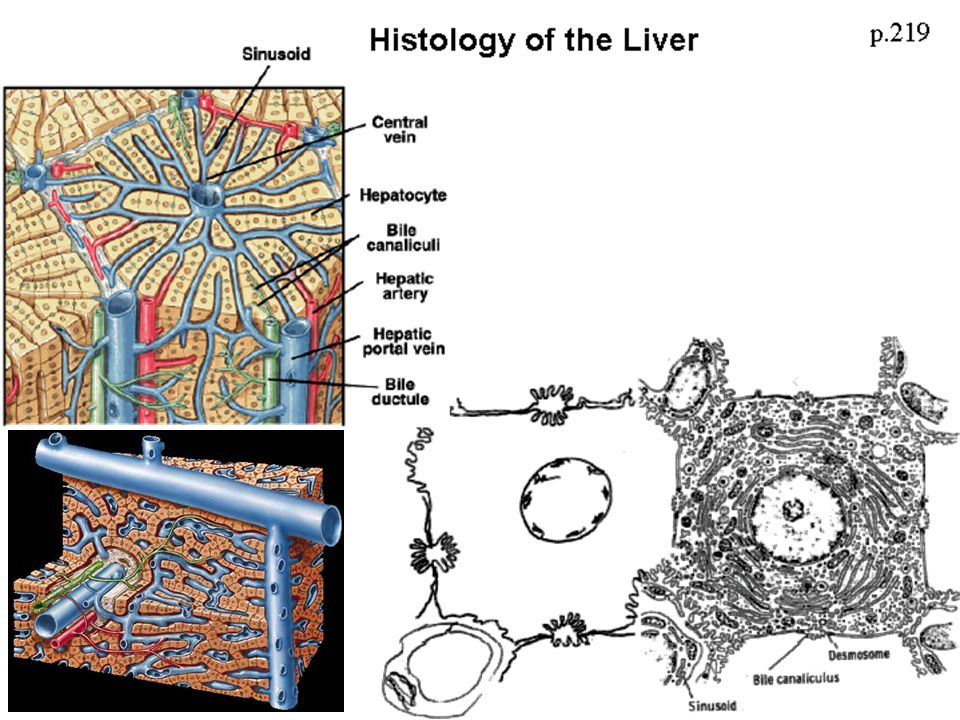
A secreção exócrina hepática

72
A secreção exócrina hepática: a bile
Bile is a digestive fluid secreted by the liver and stored in the gallbladder which normally is released into the duodenum portion of the small intestine through the sphincter of Oddi. Bile, released after a meal containing fats, aids in absorption and digestion of the fat.

73
Veja mais em: http://mcb. berkeley

74
Função digestiva da bile: sais biliares
extraído de: Vander, Sherman & Luciano, 2002

75
Sais biliares e a emulsificação das gorduras
extraído de: Vander, Sherman & Luciano, 2002

76
Sais biliares e a emulsificação das gorduras:
a formação das micelas para a digestão pela lipase pancreática MICELA extraído de: Vander, Sherman & Luciano, 2002

77
Resumo da emulsificação, digestão e absorção de gordura
extraído de: Vander, Sherman & Luciano, 2002

78
A circulação êntero-hepática dos sais biliares
extraído de: Vander, Sherman & Luciano, 2002

79
REGULAÇÃO DA SECREÇÃO BILIAR
Produção de bile + circulação êntero-hepática dos sais biliares e esvaziamento da vesícula biliar e relaxamento de Oddi pela CCK ? ? extraído de: Vander, Sherman & Luciano, 2002

80
A secreção exócrina intestinal: mucosa e hidroeletrolítica

81
Enzimas do intestino delgado:
enzimas da borda-em-escova (constitucionais) para a digestão final de proteínas e carbohidratos extraído de: Vander, Sherman & Luciano, 2002
 para a digestão final de proteínas e carbohidratos. extraído de: Vander, Sherman & Luciano,")
82
Enzimas do intestino delgado:
enzimas da borda-em-escova (constitucionais) para a digestão final de proteínas e carbohidratos
 para a digestão final de proteínas e carbohidratos.")
83
A secreção exócrina intestinal: mucosa e hidroeletrolítica
LARGE INTESTINE

84
FISIOLOGIA DO SISTEMA DIGESTÓRIO
Títulos das demais apresentações disponíveis: Introdução ao estudo do sistema digestório (SD) Movimentos observados no trato gastrointestinal (TGI) Secreções do TGI Digestão e absorção dos principais nutrientes de uma dieta ideal
 Movimentos observados no trato gastrointestinal (TGI) Secreções do TGI. Digestão e absorção dos principais nutrientes de uma dieta ideal.")
Apresentações semelhantes


, e produz o suco entérico Digestão de amido, proteínas,>")


