Carregar apresentação
A apresentação está carregando. Por favor, espere

1
UNIVERSIDADE DO VALE DO ITAJAÍ CENTRO DE CIÊNCIAS DA SAÚDE CURSO DE FARMÁCIA AVALIAÇÃO TOXICOLÓGICA DE NANOPARTÍCULAS Prof. Rilton Alves de Freitas Centro e Ciência da Saúde, Universidade do Vale do Itajai, Itajai-S.C., Brasil Aula curso Second Brazilian School on Nanobiotechnology-Itajai-S.C. 4-10 de Novembro 2007 (rede de nanobiotecnologia-NANOBIOTEC

2
ASPECTOS GERAIS DE NANOPARTÍCULAS
Fatores de toxicidade de nanopartículas (Nighswonger, 1999; Oberdörster et al., 2005; Smart et al. 2005):: Como citado anteriormente a razão área superficial pela massa, pois uma grande área superficial gera partículas com uma maior área de contato com membranas células, assim como uma grande capacidade de absorção e transporte de substâncias tóxicas. O Tempo de retenção da partícula, pois quanto maior o tempo de contato da mesma com a membrana celular, maior a chance de dano. Este fator incorpora o conceito de mobilidade de partícula, ou pelo clearance ou migração para tecidos adjacentes. Reatividade ou toxicidade inerente do composto químico contido com a partícula.
:: Como citado anteriormente a razão área superficial pela massa, pois uma grande área superficial gera partículas com uma maior área de contato com membranas células, assim como uma grande capacidade de absorção e transporte de substâncias tóxicas. O Tempo de retenção da partícula, pois quanto maior o tempo de contato da mesma com a membrana celular, maior a chance de dano. Este fator incorpora o conceito de mobilidade de partícula, ou pelo clearance ou migração para tecidos adjacentes. Reatividade ou toxicidade inerente do composto químico contido com a partícula.")
3
ASPECTOS GERAIS DE NANOFIBRAS
Tamanho da fibra x respirabilidade (capacidade de penetrar na região centro-acinar do pulmão). Fibras com comprimento maior que 15 um apenas são respiráveis se apresentam uma espessura menor que 5 um. Fibras longas (>17 um) tornam difícil a fagocitose por macrófagos, promovendo uma resposta crônica que contribui para a fibrose pulmonar. Fibras curtas, menores que 7 um, são por outro lado facilmente fagocidadas, e removidas por macrófagos alveolares (DONALDSON e TRAN, 2004; YE et al., 1999; DONALDSON et al., 1992). A Biopersistência: partículas longas são dificilmente fagocitadas por macrófagos. Fibras biosolúveis são capazes de se difundir reduzindo e serem fagocitadas mas facilmente, gerando uma baixa biopersistência e reduzindo assim a toxicidade de fibras longas longas (maiores que 20 um). Reatividade ou toxicidade inerente, dependendo basicamente dos componentes químicos da nanopartículas.
. Fibras com comprimento maior que 15 um apenas são respiráveis se apresentam uma espessura menor que 5 um. Fibras longas (>17 um) tornam difícil a fagocitose por macrófagos, promovendo uma resposta crônica que contribui para a fibrose pulmonar. Fibras curtas, menores que 7 um, são por outro lado facilmente fagocidadas, e removidas por macrófagos alveolares (DONALDSON e TRAN, 2004; YE et al., 1999; DONALDSON et al., 1992). A Biopersistência: partículas longas são dificilmente fagocitadas por macrófagos. Fibras biosolúveis são capazes de se difundir reduzindo e serem fagocitadas mas facilmente, gerando uma baixa biopersistência e reduzindo assim a toxicidade de fibras longas longas (maiores que 20 um). Reatividade ou toxicidade inerente, dependendo basicamente dos componentes químicos da nanopartículas.")
4
REGULAMENTAÇÃO DA NANOTOXICOLOGIA
AVALIAÇÃO COMPLETA DO RISCO DE PRODUÇÃO DE NANOPARTÍCULAS (Friedrichs, Schulte, 2007) Identificação do risco Caracterização do risco Avaliação do risco Prevenção e controle o risco Avaliação das medidas de controle do Obs:. Royal Society and the Royal Academy of Engineering of UK government, Recommend nanoparticulate materials to be treated as new substances.
 Identificação do risco. Caracterização do risco. Avaliação do risco. Prevenção e controle o risco. Avaliação das medidas de controle do. Obs:. Royal Society and the Royal Academy of Engineering of UK government, Recommend nanoparticulate materials to be treated as new substances.")
5
Tempo para desenvolver um medicamento
a selecionados Invenção e desenvolvimento Testes pré-clínicos (testes laboratoriais em animais) Fase I – 20 a 80 voluntários saudáveis para determinar segurança e dosagem 250 entram em teste pré-clínico Fase II – 100 a 300 voluntários para determinar eficácia e efeitos colaterais Fase III – a pacientes voluntários para monitorar reações adversas em uso de longa duração 5 entram em testes clínicos Aprovação do Governo Fase IV – Teste adicional pós-comercialização Anos 2 4 6 8 10 12 14 15 Apenas 1 chega ao mercado Patente solicitada Patente concedida
 Fase I – 20 a 80 voluntários saudáveis para determinar segurança e dosagem. 250 entram em teste pré-clínico. Fase II – 100 a 300 voluntários para determinar eficácia e efeitos colaterais. Fase III – a pacientes voluntários para monitorar reações adversas em uso de longa duração. 5 entram. em testes. clínicos. Aprovação do Governo. Fase IV – Teste adicional pós-comercialização. Anos Apenas 1 chega ao mercado. Patente. solicitada. Patente. concedida.")
6
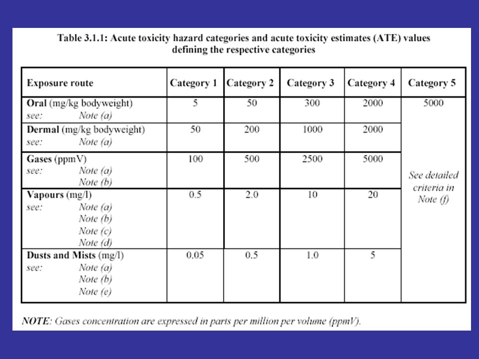
Classificação toxicológica
Avaliação toxicológica – Estudos Pré- clínicos Estudos Agudos DL50 Oral DL50 Dérmica CL50 Inalatória Irritação / Corrosão Ocular Irritação / Corrosão Dérmica Sensibilização Cutânea Avaliação de perigo Classificação toxicológica

7
Avaliação toxicológica
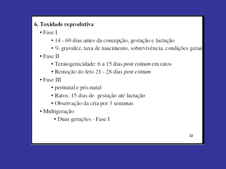
Estudos sobre mutagenicidade Mutação gênica (procariontes) Mutação cromossômica (eucariontes) Estudos sobre carcinogenicidade Camundongo (18 meses) Rato (24 meses) Proibição do Registro Estudos sobre teratogenicidade Coelho Rato
 Mutação cromossômica (eucariontes) Estudos sobre carcinogenicidade. Camundongo (18 meses) Rato (24 meses) Proibição do. Registro. Estudos sobre teratogenicidade. Coelho. Rato.")
8
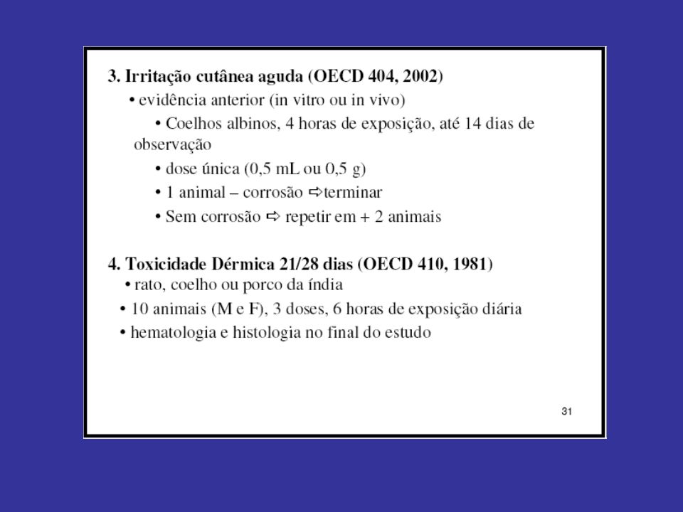
Avaliação toxicológica
Estudos de curto prazo Roedor (90 dias) Não roedor (1 ano) Estudos de longo prazo e carcino Camundongo (18 meses) Rato (24 meses) NOAEL Estudos de neurotoxicidade Estudos de reprodução e prole Estudos dos efeitos teratogênicos
 Não roedor (1 ano) Estudos de longo prazo e carcino. Camundongo (18 meses) Rato (24 meses) NOAEL. Estudos de neurotoxicidade. Estudos de reprodução e prole. Estudos dos efeitos teratogênicos.")
9
Definições importantes
Ingestão Diária Aceitável (IDA) - quantidade máxima que, ingerida diariamente durante toda a vida, parece não oferecer risco apreciável à saúde, à luz dos conhecimentos atuais
 - quantidade máxima que, ingerida diariamente durante toda a vida, parece não oferecer risco apreciável à saúde, à luz dos conhecimentos atuais.")
10
Avaliação toxicológica
Dose 3X NOAEL Dose 2X Dose X Controle

11
Avaliação toxicológica
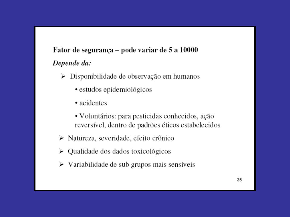
NOAEL IDA = NOAEL Fator de incerteza Fatores de incerteza

13
TESTES PRÉ-CLÍNICOS: MÉTODOS TOXICOLOGICOS “IN VITRO”
Azul de tripan

14
QUANTIFICAÇÃO DE CRESCIMENTO
MTT (3-(4,5-dimethylthiazole-2-yl)-2,5-diphenyl tetrazolium bromide)
-2,5-diphenyl tetrazolium bromide)")
15
Alaranjado de Acridina
Apoptosis Necrosis Staining with acridine orange and propidium iodide makes it possible to discriminate between different cells. Late apoptotic cells only stain with acridine orange, that can pass the still intact membrane. It stains the DNA, making it possible to observe the condensed nature of the chromatin. Very late apoptotic cells lose their membrane integrity and stain with PI giving a red colour. The condensed nature of the chromatin can still be observed. For necrotic cells also the DNA is stained by both stains. However, since the DNA is not condensed a diffuse colour all over the nucleus is observed. Since cells lose their membrane integrity very rapidly all necrotic cells stain red. In practise it is very difficult and subjective to discriminate the different cells. However, lately better dyes have become available. AO stains DNA Small intense spots Diffuse spots

16
Processos necróticos: Lactato desidrogenase
NAD NADH LACTATO PIRUVATO

17
QUANTIFICAÇÃO APOPTOSE
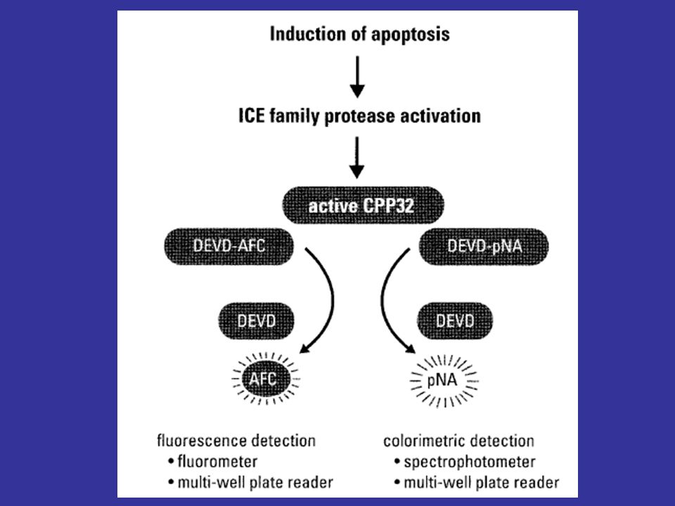
Processos terminais Nucleases: Fragmentação do DNA Morfologia (nuclear) Processos iniciais Fosfatidilserina Caspases There are different methods to measure apoptosis. Late apoptotic cells can be measured by the fragmented DNA. Nucleases cut the DNA between the nucleosomes giving a ladder-like pattern on a DNA gel. Furthermore the strand breaks can be detected through specific end-labelling tests. Furthermore these cells can be detected by their morphology. Chromatin is condensed at the borders of the nucleus and the membrane of the cells blebs and the cell shrinks. Early apoptotic cells can be detected by the fact that early in the process a specific lipid, phosphatidyl serine, which normally is on the cytoplasmic site of the membrane is externalised. This can be detected. Furthermore a specific family of proteases called caspases become active early in apoptosis. This enzyme activity can be measured
 Processos iniciais. Fosfatidilserina. Caspases. There are different methods to measure apoptosis. Late apoptotic cells can be measured by the fragmented DNA. Nucleases cut the DNA between the nucleosomes giving a ladder-like pattern on a DNA gel. Furthermore the strand breaks can be detected through specific end-labelling tests. Furthermore these cells can be detected by their morphology. Chromatin is condensed at the borders of the nucleus and the membrane of the cells blebs and the cell shrinks. Early apoptotic cells can be detected by the fact that early in the process a specific lipid, phosphatidyl serine, which normally is on the cytoplasmic site of the membrane is externalised. This can be detected. Furthermore a specific family of proteases called caspases become active early in apoptosis. This enzyme activity can be measured.")
18
BROMETO DE ETIDIO ALARANJADO DE ACRIDINA
DAPI TUNEL/IODETO PROPIDIO

19
Células saudáveis Células vitais Células intactas Células mortas
Membrana polarizada DAPI Sistema de transporte ativo (Azul 460 nm) Células vitais Membrana polarizada BROMETO DE ETIDIO Sem sistema de transporte ativo (Vermelho 602 nm) Células intactas Membrana despolarizada BROMETO DE ETIDIO Células mortas Membrana despolarizada BROMETO DE ETIDIO Sem sistema de transporte ativo IODETO PROPIDIO (Vermelho 603 nm)
 Células vitais. Membrana polarizada BROMETO DE ETIDIO. Sem sistema de transporte ativo (Vermelho 602 nm) Células intactas. Membrana despolarizada BROMETO DE ETIDIO. Células mortas. Membrana despolarizada BROMETO DE ETIDIO. Sem sistema de transporte ativo IODETO PROPIDIO (Vermelho 603 nm)")
20
- - - Annexine-FITC early + PI late normal
A method to detect early apoptotic cells is based on the externalisation of phosphatidyl choline. Phosphatidyl serine is a phospho-lipid normally only found on the cytoplasmic side of the plasma membrane. This is caused by an active floppase. Working against this is a scramblase that randomly flips the lipid. Upon apoptosis the floppase action is stopped and phosphatidyl serine is exposed on the outside of the membrane. In the body this is a signal for specialised macrophages to phagocytose the cell. PS on the outside can be detected by binding of annexine labelled with a green fluorescent dye FITC. At the same time PI is added. Thus cells that only stain green are early apoptotic and cells that stain red and greed are late apoptotic also called secondary necrotic. It is not clear whether cells can actually reach the inside PS. Furthermore, it is not sure whether necrotic cells also externalise the PS, but since this is an active process probably not. late normal

21
Citometria de Fluxo Espalhamento (Forma, Granulos) size
Fluorescence Verde (FITC) Fluorescence Vermelho (PI) The fluorescence of individual cells can be measured using flow-cytometry. In a flow cytometer a flow of liquid suspended cells is generated in a measurement chamber. A laser beam crosses this flow and if a cell passes with the flow the light is scattered. The scattered light is measured under a small angle, the forward scatter, and a large angle, the side scatter. The FSc is a measure for the size of the cell, while the side scatter is a measure of the granularity of the cell and roughness of the cell membrane. Apoptotic cells have a more blebbed membrane are smaller and are more granular than viable cells. If cells are stained with a fluorescent dyes the fluorescence can be measured for each cell.
 Fluorescence Vermelho (PI) The fluorescence of individual cells can be measured using flow-cytometry. In a flow cytometer a flow of liquid suspended cells is generated in a measurement chamber. A laser beam crosses this flow and if a cell passes with the flow the light is scattered. The scattered light is measured under a small angle, the forward scatter, and a large angle, the side scatter. The FSc is a measure for the size of the cell, while the side scatter is a measure of the granularity of the cell and roughness of the cell membrane. Apoptotic cells have a more blebbed membrane are smaller and are more granular than viable cells. If cells are stained with a fluorescent dyes the fluorescence can be measured for each cell.")
22
Dados Típicos Side scatter Side scatter Espalhamento lateral
Forward scatter Espalhamento frontal Side scatter Espalhamento lateral Side scatter Here we see so called contour plots for cells from a steady state continuous culture at a low dilution and a high dilution rate. At the low dilution rate the viability is low and many cells are apoptotic as can be seen from the population with a higher side scatter and smaller forward scatter. Furthermore, we see a kind of band towards the cell debris in the corner. Forward scatter

23
PI vs. Annexin-FITC) PI Late apoptotic Necrotic ? Early apoptotic
Viable Here we see cells from a batch culture in the death phase. On the x-axis the amount of green fluorescence or annexin binding and on the y-axis the red fluorescence or the amount of PI taken up.As we can see there is a population of viable cells at low red and green fluorescence, a population of early apoptotic cells at high green but low red fluorescence and a population of late apoptotic cells at high red and green fluorescence. It is up to now unclear what the population at high red and low green fluorescence represents. They could be necrotic cells or late apoptotic cells for which the annexin has been cut from the cells. Annexine-FITC

24
Ciclo celular + corantes
Propidium iodide = DNA BRdU = Sintese DNA S=DNA synthesis M=cell divides G = gap G1 S G2 M Apoptose Another type of measurement is the cell-cycle measurement. Here we see the cell cycle. In the M-phase the cell divides, while in the S-phase DNA is synthesised. In between are the gap phases G1 and G2. Cells are first incubated with Bromo-desoxy-Uridine for one hour at 37 C. BrdU is an analogue of thymidine that is build into the DNA of DNA-synthesising cells. Next cells are harvested and fixed with ethanol. After denaturation of the DNA cells are stained with propidium iodide that intercalates in the DNA and a green fluorescent antibody against BrDU. This gives the following results: cells that are in G1 have a single DNA content and red fluorescence. Cells in G2 have twice the amount of DNA and thus about twice red fluorescence and finally S-phase cells have a DNA content and red fluorescence in between the G1 and G2/M cells. Since S-phase cells synthesise DNA they also fluorescence green. In late apoptotic cells the DNA is cut into fragments, which upon ethanol treatment diffuse out of the cell. This causes a red fluorescence for these cells even lower than that of the G1 cells, the so called sub-G1 peek. Losing DNA

25
S S O O G2/M G2/M G1 G1 Green Green Red fluorescence Red fluorescence
Here we see a flow-cytometric measurement for the cell-cycle analysis. Each dot is a cell positioned according to its red and green fluorescence. The right figure is a sample from a high dilution rate chemostat meaning a viable culture. Here is the G1. At twice the red fluorescence the G2/M and at intermittent red and high green fluorescence the S-phase cells. The apoptotic cells are expected on the left. If we look at a sample from a low dilution rate low viability chemostat we see a significant apoptotic population and furthermore that the relative amount of S-phase cells has decreased. Red fluorescence Lower dilution rate Higher death Higher dilution rate Lower death

27
WESTERN BLOT OU IMMUNOBLOT
CASPASE -3 WESTERN BLOT OU IMMUNOBLOT

28
FRAGMENTAÇÃO DNA

29
MÉTODOS DE AVALIAÇÃO DA BIODISPONIBILIDADE “IN VITRO” DE NANOPARTÍCULAS
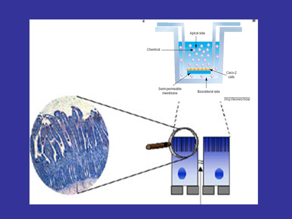
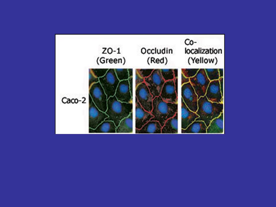
Células Caco-2 culture dish insert Basolateral medium apical medium Caco-2 cell monolayer Caco-2 cells were used as a model for small intestine. Caco-2 cells are derived from human colon cancer cells and are usually grown as monolayers in a transwell such that there is an apical side and a basolateral side to the cells. DHB was added to the apical side to mimic grapefruit ingestion. Upon differentiation resemble small intestinal enterocytes. Lee ren in our lab has shown a Vitamin D analog induces CYP3a4 expression. Therefore it is a good in vitro model to study intestinal CYP3A4

30
Permeação aparente (Papp)
células/mL Matriz extracelular Célula Caco-2 Meio E-MEM 10% SFB Incubação 14 – 28 dias 37oC/5%CO2 TEER 350 *cm2 Baso Lateral Porção apical Tempos 0 – 24 horas Intervalo 2h

31
CLASSIFICAÇÃO BIOFARMACÊUTICA
Solubilidade e Permeabilidade são os parâmetros chaves que controlam a absorção de fármacos (AMIDON, 1995) Classe I: Fármacos de alta solubilidade e alta permeabilidade; Classe II: Fármacos de baixa solubilidade e alta permeabilidade; Classe III: Fármacos de alta solubilidade e baixa permeabilidade; Classe IV: Fármacos de baixa solubilidade e baixa permeabilidade
 Classe I: Fármacos de alta solubilidade e alta permeabilidade; Classe II: Fármacos de baixa solubilidade e alta permeabilidade; Classe III: Fármacos de alta solubilidade e baixa permeabilidade; Classe IV: Fármacos de baixa solubilidade e baixa permeabilidade.")
32
Caco-2 cells were used as a model for small intestine
Caco-2 cells were used as a model for small intestine. Caco-2 cells are derived from human colon cancer cells and are usually grown as monolayers in a transwell such that there is an apical side and a basolateral side to the cells. DHB was added to the apical side to mimic grapefruit ingestion. Upon differentiation resemble small intestinal enterocytes. Lee ren in our lab has shown a Vitamin D analog induces CYP3a4 expression. Therefore it is a good in vitro model to study intestinal CYP3A4

36
Manitol > 300 cm < 300 cm2 TEER: Resistência Elétrica Transepitelial

37
Apical enterocito Basal

38
APICAL * enterocito CYP3A4 BASAL

39
APICAL enterocito * X CYP3A4 BASAL

40
APICAL enterocito * BASAL

41
APICAL Pgp OATP enterocito CYP3A4 BASAL

42
Razão dose/solubilidade
BIODISPONIBILIDADE Conc. Basal tg TEMPO (h) Classificação do fármaco segundo o Sistema de Classificação Biofarmacêutico. Classe Papp Razão dose/solubilidade 1 Altamente solúveis e altamente permeáveis > 10-5 < ou = 0,5 2 Baixa solubilidade e alta permeabilidade > 0,5 3 Alta solubilidade e baixa permeabiliade < 2 x 10⁻⁶ 4 baixa solubilidade e baixa permeabilidade > 1
 Classificação do fármaco segundo o Sistema de Classificação Biofarmacêutico. Classe. Papp. Razão dose/solubilidade. 1 Altamente solúveis e altamente permeáveis. > < ou = 0,5. 2 Baixa solubilidade e alta permeabilidade. > 0,5. 3 Alta solubilidade e baixa permeabiliade. < 2 x baixa solubilidade e baixa permeabilidade. > 1.")
43
Papp > 10-6 cm/s: 100% absorvido
10-7 < Papp < 10-6 cm/s: 1 – 99% absorção Papp < 10-7 cm/s: < 1 % absorção Bioequivalência: Alta permeabilidade Papp > 10-5 cm/s Baixa permeabilidade Papp < 10-5 cm/s

44
TESTES PRÉ-CLÍNICOS: TESTES TOXICOLÓGICOS “IN VIVO”

49
4. MUTAGÊNESE (GENOTOXICIDADE) E CARCINOGÊNESE
Acridinas = intercalantes Levam a mudança de fase de leitura Dímero de pirimidina Lesão mais comum do UV curto (fotoliase) Nem sempre a droga é mutagênica Aflatoxina do fungo do amendoim é ativada perto do núcleo
 Nem sempre a droga é mutagênica. Aflatoxina do fungo do amendoim. é ativada perto do núcleo.")
50
Reparo de erros de pareamento “Mismatch repair” Expansão de repetições
Huntintina expande de 6-31 rep. para 36-82 Teste de AMES 10 bilhões de Salmonella his- Uma reversão de mutação torna-as his+

51
GENOTOXICIDADE Lavar com PBS gelado Remoção das epífises Camundongo
1) Contagem câmara de Neubauer 2) Viabilidade 85% 3) cel/mL Camundongo Fêmur Lâminas Jateadas Agarose 1% Incubar 1h 37C/5%CO2 Controles DMSO MMS ou H2O2 Agarose Low melting 0,75%
 Contagem. câmara de Neubauer. 2) Viabilidade 85% 3) cel/mL. Camundongo. Fêmur. Lâminas Jateadas. Agarose. 1% Incubar 1h 37C/5%CO2. Controles. DMSO. MMS ou H2O2. Agarose. Low melting. 0,75%")
52
Tampão de corrida 25 V 300 mA 30 min Tampão de Neutralização Brometo de etídeo Tampão de lise DMSO: Núcleo íntegro (controle NEGATIVO de Genotoxicidade) MMS: Fragmentação nuclear (controle POSITIVO de Genotoxicidade)
 MMS: Fragmentação nuclear (controle POSITIVO de Genotoxicidade)")
53
Método de classificação de Kobayashi et al. (1995); Miyamae et al
1)sem cauda 2) cometas com pequenas caudas (caudas com comprimento < 25% da cabeça) 3) Cometas com caudas de tamanho médio (caudas com comprimento entre 25% e 100% da cabeça) 4) Cometas com caudas longas (comprimento da cauda > que a cabeça) 5) Cometas mal definidos ou com cabeça pequena.
sem cauda. 2) cometas com pequenas caudas (caudas com comprimento < 25% da cabeça) 3) Cometas com caudas de tamanho médio (caudas com comprimento entre 25% e 100% da cabeça) 4) Cometas com caudas longas (comprimento da cauda > que a cabeça) 5) Cometas mal definidos ou com cabeça pequena.")
Apresentações semelhantes


>")


