Carregar apresentação
A apresentação está carregando. Por favor, espere

1
Anatomia Sistema Respiratório II Parte II
Direitos Reservados ® Centro de Estudos Avançados em Saúde® CEAS Anatomia Sistema Respiratório II Parte II Prof Danillo Barbosa

2
Bibliografia recomendada (livros-textos)
“Fisiologia” Constanzo, 2004, 2ª Ed. (Ed. Elsevier) “Fundamentos de Fisiologia”, Berne et al, 2006, 4ª Ed.(Ed. Elsevier) “Fisiologia” Berne et al., 2004 (Ed. Elsevier) “Tratado de Fisiologia Médica” Guyton & Hall, 2006, 11ª Ed. (Ed. Elsevier) “Fundamentos de Fisiologia Médica” Johnson, 2003 (Ed. Guanabara Koogan) “Fisiologia: texto e atlas” Silbernagl e Despopoulos, 2003 (Ed. Artmed) Prof Danillo Barbosa
 Fundamentos de Fisiologia , Berne et al, 2006, 4ª Ed.(Ed. Elsevier) Fisiologia Berne et al., 2004 (Ed. Elsevier) Tratado de Fisiologia Médica Guyton & Hall, 2006, 11ª Ed. (Ed. Elsevier) Fundamentos de Fisiologia Médica Johnson, 2003 (Ed. Guanabara Koogan) Fisiologia: texto e atlas Silbernagl e Despopoulos, 2003 (Ed. Artmed) Prof Danillo Barbosa.")
3
VEJA TEXTOS COM ANIMAÇÕES ONLINE
Section 4 - Respiratory Physiology Section 4 Ch 1 pg 16 Alveolarization and Alveolar Surface Area Section 4 Ch 1 pg 18 Measurement of Lung and Ventilation Volumes Section 4 Ch 1 pg 22 Circulatory Transport of O2 and CO2 Section 4 Ch 2 pg 4 Respiratory Mechanics: Identification of Forces Section 4 Ch 2 pg 11 The Diaphragm Section 4 Ch 2 pg 12 External Intercostal Muscles Section 4 Ch 2 pg 15 Expiratory Muscle Groups Section 4 Ch 2 pg 21 The Opposing Force of Pulmonary Elastance or Compliance Section 4 Ch 2 pg 23 Lung Compliance Curve Section 4 Ch 2 pg 24 Chest Cage Compliance Curve Section 4 Ch 2 pg 25 Combined Lung-Chest Wall Compliance: The Relaxation Curve Section 4 Ch 2 pg 31 Elastic and Collagen Fibers of the Lung Section 4 Ch 2 pg 35 Alveolar Instability with Constant Surface Tension Section 4 Ch 2 pg 37 Surface Tension in Lung Lavage Fluids Section 4 Ch 2 pg 40 asid Interdependence Section 4 Ch 2 pg 54 Physical Factors that Affect Airway Resistance Section 4 Ch 2 pg 54 Physical Factors that Affect Airway Resistance: enlargement of the airway lumen Section 4 Ch 3 pg 21 Alveolar Ventilation Section 4 Ch 4 pg 7 Pulmonary Circulation as a Blood Filter Section 4 Ch 4 pg 8 Reservoir of Blood for Left Ventricle Section 4 Ch 4 pg 15 Factors Affecting Lung Pressures: Alveolar Pressure Section 4 Ch 4 pg 17 Measurement of Pulmonary Vascular Pressures with Swan-Ganz Catheter Section 4 Ch 5 pg 5 Solubilities of Respiratory Gases Section 4 Ch 5 pg 21 Significance of Shifts in the Oxy-Hb Dissociation Section 4 Ch 7 pg 29 Hypoxic Hypoxemia with Emphysema Section 4 Ch 8 pg 19 Flow Volume Loops in Detection of Pulmonary Disorders Fisiologia Humana Prof.Ms.Danillo Barbosa

5
Prof.Ms.Danillo Barbosa
Fisiologia Humana Prof.Ms.Danillo Barbosa

6
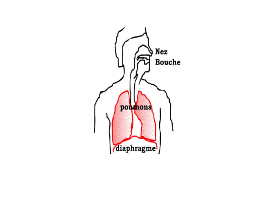
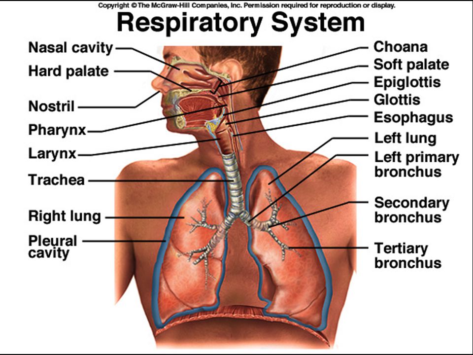
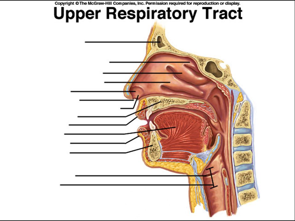
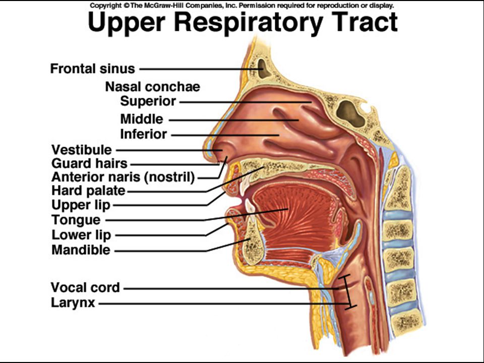
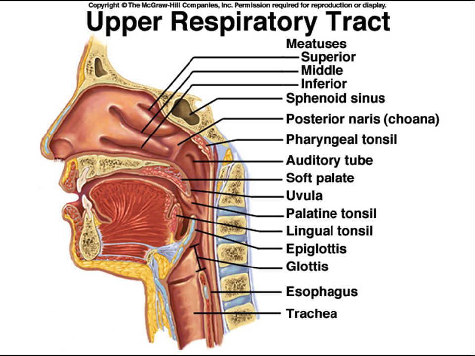
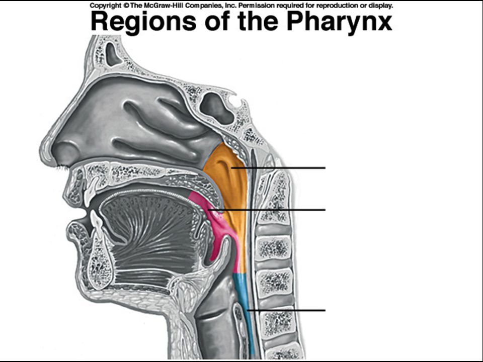
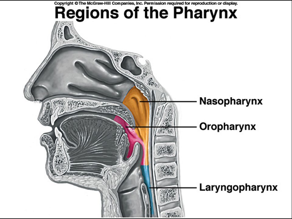
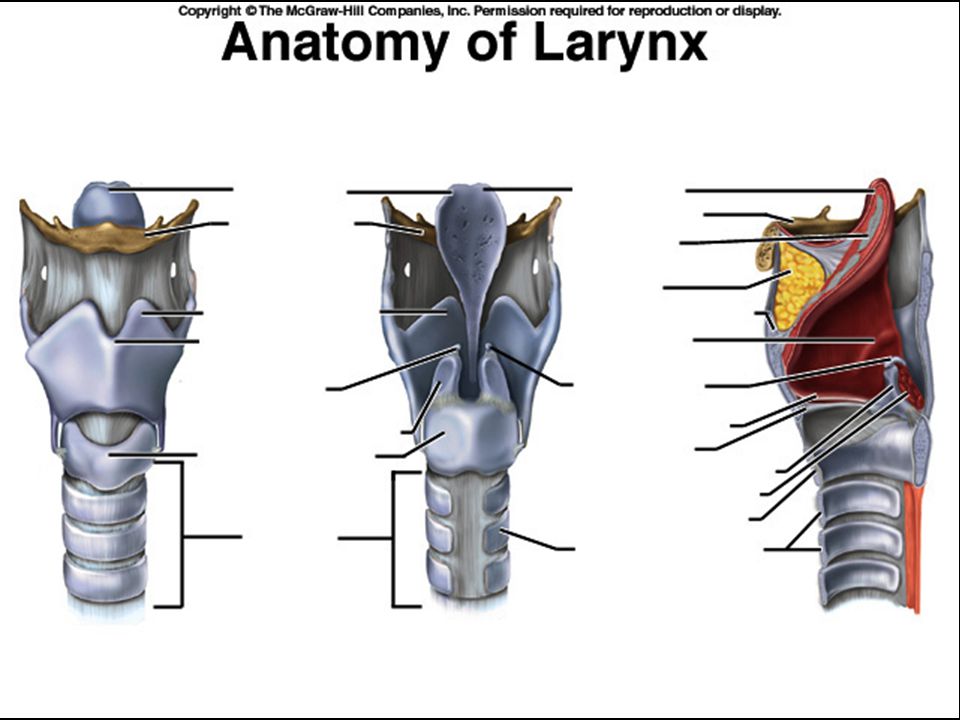
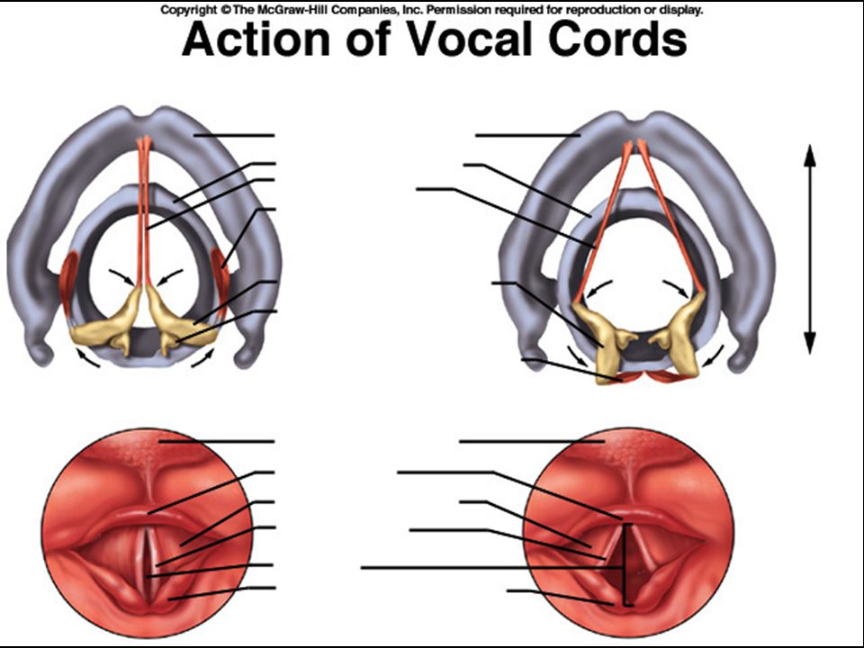
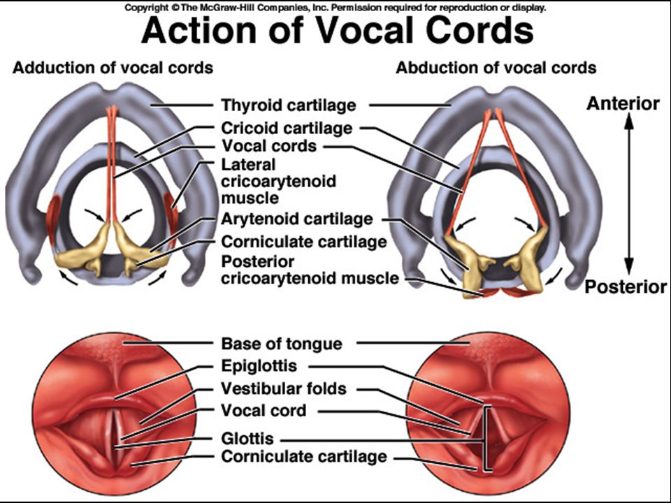
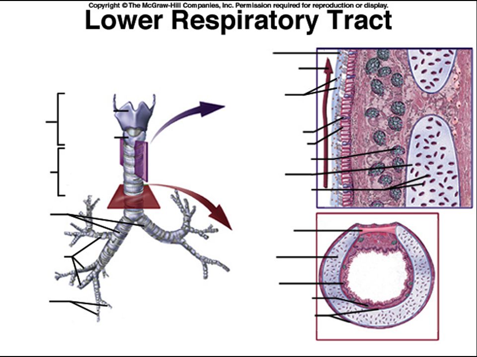
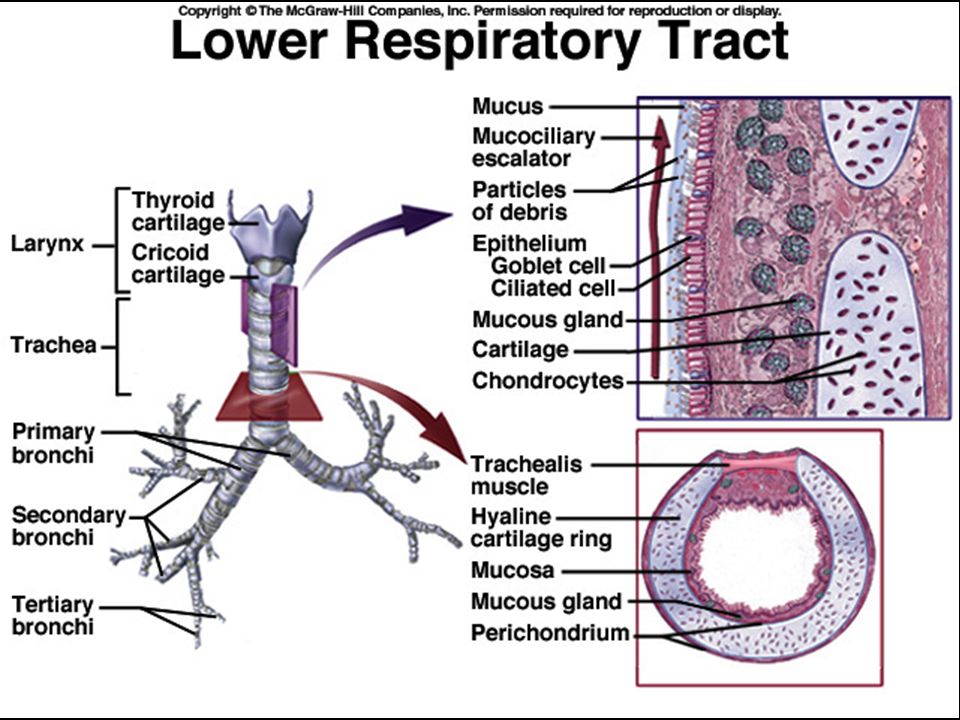
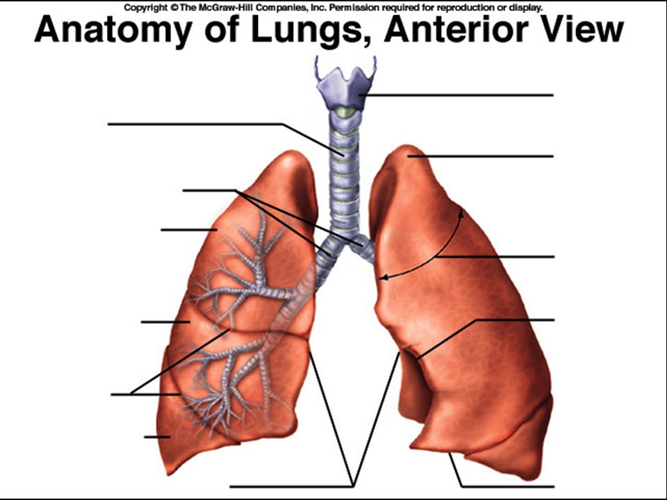
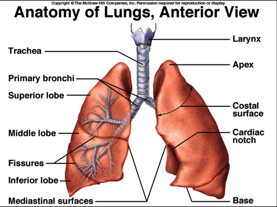
Anatomia Sistema Respiratório
Fisiologia Humana Prof.Ms.Danillo Barbosa

7
DIAFRAGMA

8
DIAFRAGMA site original (retirado):
:")
9
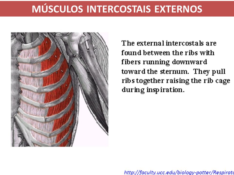
MÚSCULOS INTERCOSTAIS EXTERNOS
10
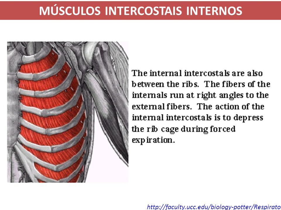
MÚSCULOS INTERCOSTAIS INTERNOS
11
MÚSCULOS ABDOMINAIS DA EXALAÇÃO
site original (retirado):
:")
28
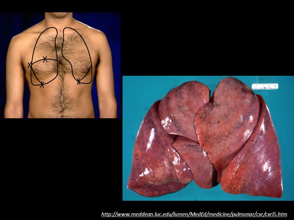
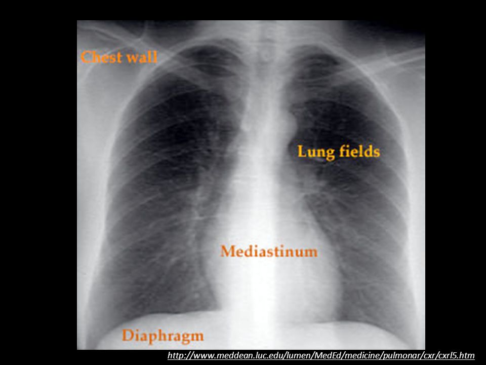
http://www. meddean. luc. edu/lumen/MedEd/medicine/pulmonar/cxr/cxrl5
29
http://www. meddean. luc. edu/lumen/MedEd/medicine/pulmonar/cxr/cxrl5

30
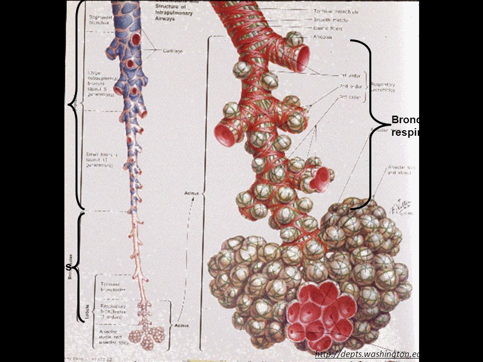
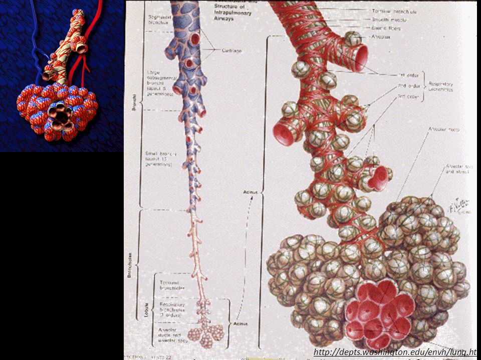
brônquios bronquíolos Bronquíolos respiratórios
31
These two scanning electron micrographs show the organization of the pulmonary acinus. This micrograph is of a cast of two terminal bronchioles, the short respiratory, or transitional, bronchioles and all of the alveolar air spaces supplied by those bronchioles. SETA: bronquíolo respiratório.

33
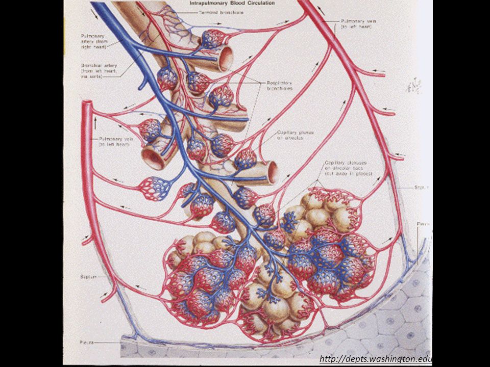
Resin cast of pulmonary arteries and airways
Resin cast of pulmonary veins and airways

35
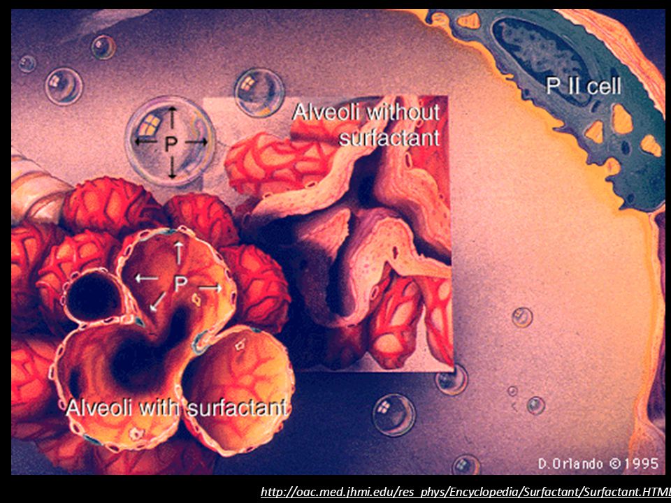
http://oac. med. jhmi. edu/res_phys/Encyclopedia/Surfactant/Surfactant
36
Prof.Ms.Danillo Barbosa
Edema Pulmonar Fisiologia Humana Prof.Ms.Danillo Barbosa

38
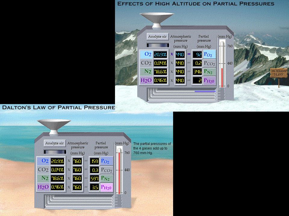
Pressão atmosférica: 760 mm Hg

42
Prof.Ms.Danillo Barbosa
Fisiologia Humana Prof.Ms.Danillo Barbosa

46
http://www. meddean. luc. edu/lumen/MedEd/medicine/pulmonar/cxr/cxrl5
47
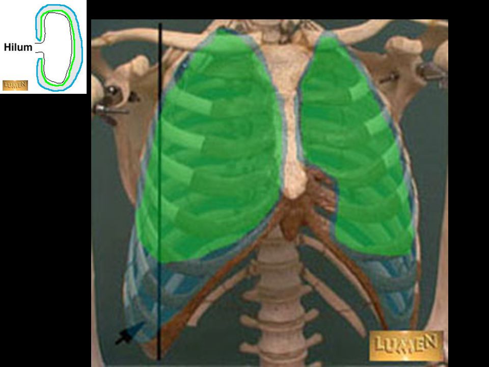
The pleural cavities are closed sacs enveloping each lung
The pleural cavities are closed sacs enveloping each lung. Each cavity comprises a visceral layer (green) and a parietal layer (blue).
 and a parietal layer (blue).")
49
Pneumothorax A pneumothorax is a condition in which air has entered and expanded the normally closed pleural space, driving pleural pressure up toward atmospheric pressure, and resulting in partial or complete collapse of the lung. When pleural pressure approaches zero, the lung and chest wall both move toward the equilibrium positions they would assume in the absence of any external pressures-- the lung collapses and the chest wall springs out. A pneumothorax may be induced by a break in either the parietal pleura (e.g., from trauma, needle or catheter insertion) or in the visceral pleura (e.g., from rupture of a subpleural air pocket or necrosis of lung adjacent to the pleura). The right side of this patient's thoracic cavity (viewer's left) shows a darker area where the lung should be. Note the expanded chest wall and the collapsed lung.
 or in the visceral pleura (e.g., from rupture of a subpleural air pocket or necrosis of lung adjacent to the pleura). The right side of this patient s thoracic cavity (viewer s left) shows a darker area where the lung should be. Note the expanded chest wall and the collapsed lung.")
50
veja aqui texto e animação online
O CICLO RESPIRATÓRIO veja aqui texto e animação online

51
veja aqui texto e animação online
O CICLO RESPIRATÓRIO veja aqui texto e animação online

52
veja aqui texto e animação online
O CICLO RESPIRATÓRIO veja aqui texto e animação online

53
veja aqui texto e animação online
O CICLO RESPIRATÓRIO veja aqui texto e animação online

54
veja aqui texto e animação online
O CICLO RESPIRATÓRIO veja aqui texto e animação online

55
VOLUMES PULMONARES

56
volume de ar inspirado e expirado em cada ciclo ventilatório normal.
Volume corrente (VT): volume de ar inspirado e expirado em cada ciclo ventilatório normal. (~500ml) VT: “Tidal Volume”
: volume de ar inspirado e expirado em cada ciclo ventilatório normal. (~500ml) VT: Tidal Volume")
57
Volume de reserva inspiratória (VRI):
volume de ar que ainda pode ser inspirado ao final da inspiração do volume corrente normal (~3.000ml) Volume corrente (VT):
 Volume corrente. (VT):")
58
Volume de reserva inspiratória (VRI):
Volume corrente (VT): Volume de reserva expiratória (VRE): volume de ar que, por meio de uma expiração forçada, ainda pode ser exalado ao final da expiração do volume corrente normal (~1.100ml)
: Volume de reserva expiratória (VRE): volume de ar que, por meio de uma expiração forçada, ainda pode ser exalado ao final da expiração do volume corrente normal. (~1.100ml)")
59
Volume de reserva inspiratória (VRI):
Volume corrente (VT): Volume de reserva expiratória (VRE): Volume residual (VR): volume de ar que permanece nos pulmões mesmo ao final da mais vigorosa das expirações (~1.200ml). Não pode ser medido por espirometria
: Volume de reserva expiratória (VRE): Volume residual (VR): volume de ar que permanece nos pulmões mesmo ao final da mais vigorosa das expirações (~1.200ml). Não pode ser medido por espirometria.")
60
CAPACIDADES PULMONARES
VOLUMES PULMONARES CAPACIDADES PULMONARES

61
Capacidade inspiratória (CI): VT + VRI
Essa quantidade de ar é aquela que uma pessoa pode inspirar, partindo do nível expiratório basal e enchendo ao máximo os pulmões (~3.500ml).
.")
62
Capacidade inspiratória (CI): VT + VRI
Capacidade Residual Funcional (CRF): VRE + VR Essa quantidade de ar (~2.300ml) é a que permanece nos pulmões ao final da expiração normal. Não pode ser calculada por espirometria
: VRE + VR. Essa quantidade de ar (~2.300ml) é a que permanece nos pulmões ao final da expiração normal. Não pode ser calculada por espirometria.")
63
Capacidade inspiratória (CI): VT + VRI
Capacidade Vital (CV): VRI + VT + VRE É a maior quantidade de ar que uma pessoa pode expelir dos pulmões após tê-los enchido ao máximo e, em seguida, expirado completamente (~4.600ml) Capacidade Residual Funcional (CRF): VRE + VR
: VRI + VT + VRE. É a maior quantidade de ar que uma pessoa pode expelir dos pulmões após tê-los enchido ao máximo e, em seguida, expirado completamente (~4.600ml) Capacidade Residual Funcional (CRF): VRE + VR.")
64
Capacidade inspiratória (CI): VT + VRI
Capacidade Vital (CV): VRI + VT + VRE Capacidade Pulmonar Total (CPT): VRI + VT + VRE + RV É o maior volume que os pulmões podem alcançar (~5.800ml) ao final do maior esforço inspiratório possível. Capacidade Residual Funcional (CRF): VRE + VR
: VRI + VT + VRE. Capacidade Pulmonar Total (CPT): VRI + VT + VRE + RV. É o maior volume que os pulmões podem alcançar (~5.800ml) ao final do maior esforço inspiratório possível. Capacidade Residual Funcional (CRF): VRE + VR.")
65
FIM

Apresentações semelhantes


>")


