Carregar apresentação
A apresentação está carregando. Por favor, espere

1
Marcadores Moleculares
Rodrigo Rocha Latado

2
Marcadores fenotípicos
Características do organismo - Ex. cor da pele, cor e tipo de cabelo, presença de pelos, etc... Marcadores bioquímicos Presença ou ausência de compostos - Ex. isoenzimas, proteínas ou de polissacarídeos específicos, etc... Marcadores moleculares Presença ou ausência de sequências de DNA ou RNA - Ex. genes ou regiões não-codificantes - alteração em uma ou mais bases nitrogenadas

3
USOS Testes de paternidade Exames genéticos - aberrações cromossômicas ou SNP´s) para Humanos, animais, plantas e microorganismos Causas forenses (criminais, patenteamento)
")
4
USOS Estudos de fingerprinting (impressões digitais) - Biochips de DNA Estudos básicos de genômica e proteômica Estudos ambientais (conservação de germoplasma, avaliação da diversidade genética)
")
5
USOS Estudos evolucionários e populacional Melhoramento animal e vegetal (seleção assistida, seleção de progenitores)
 .")
6
CLASSIFICAÇÃO DE MARCADORES MOLECULARES

7
Classificação por Tipo de Técnica
Métodos sem uso de PCR RFLP VNTR Métodos com uso de PCR PCR com primers arbitrários RAPD, AP-PCR, DAF; Polimorfismo de Tamanho de Fragmento Amplificado AFLP; ISSR TRAP PCR sítio-específico CAPS, SCAR SSRs (microssatélites) TGGE, SSCP, DGGE
 TGGE, SSCP, DGGE.")
8
Classificação por Número de Cópias da Seqüência Alvo
Seqüência de poucas cópias - codificante RFLP Seqüência com cópias repetidas VNTR SSRs (microssatélites) ISSR Seqüência com número de cópias indefinido RAPD, AP-PCR, DAF; AFLP; CAPS, SCAR TRAP
 ISSR. Seqüência com número de cópias indefinido. RAPD, AP-PCR, DAF; AFLP; CAPS, SCAR. TRAP.")
9
MARCADORES MOLECULARES PARA MELHORAMENTO DE PLANTAS

10
Marcadores genéticos são unidades herdáveis simples
Sucesso no melhoramento de plantas depende da capacidade de distinguir fatores genéticos herdáveis, dos ambientais Marcadores genéticos são unidades herdáveis simples marcadores cromossoma

11
Polimorfismo de DNA é resultado do acúmulo de mutações
pontual ou inserção/deleção macro-rearranjos: translocações, inversões, deleções Ex. magnitude mutações pontuais: Gene Opaco-2 : pares de bases 14% das bases polimorfismo 26 fenômenos de Inserção/Deleção

12
Marcadores genéticos, quando associados a características de interesse, aumentam a eficiência de seleção R cromossoma

13
População de São Paulo 9.291.000 habitantes em 2.600.000 domicílios
Como achar alguém? E se fosse Tokyo?

14
Aplicações no Melhoramento
Mapeamento genômico Seleção Assistida por Marcadores - MAS Diversidade genética e filogenia Caracterização de germoplasma Identificação de acessos Seleção de genitores

15
Histórico de Marcadores
’s - ferramentas moleculares desenvolvimento de vetores de clonagem; enzimas de restrição; polimerases; ligases; Southern (1977); 2. RFLP proposto por Botstein et al. (1980) descrito para humanos 3. PCR proposto por Mullis & Faloona (1987) 4. RAPD por Rafalski et al. (1990)
; 2. RFLP proposto por Botstein et al. (1980) descrito para humanos. 3. PCR proposto por Mullis & Faloona (1987) 4. RAPD por Rafalski et al. (1990)")
16
Histórico de Marcadores
5. Microssatélites em plantas por Akkaya et al. (1992) 6. AFLP por Zabeau & Vos (1993) 7. SCAR por Paran & Michelmore (1993) 8. Cho et al. (1999) - SNPs em Arabidopsis
 6. AFLP por Zabeau & Vos (1993) 7. SCAR por Paran & Michelmore (1993) 8. Cho et al. (1999) - SNPs em Arabidopsis.")
17
DESCRIÇÃO DOS MARCADORES MOLECULARES

18
Marcadores Moleculares
RFLP RAPD, AP-PCR, DAF PCR-específico - SSR, ISSR, CAPS, SCARs AFLP SNPs

19
Restriction Fragment Length Polymorphism - RFLP
RFLP examina diferenças no tamanho de fragmentos de restrição de DNA específicos Polimorfismo deriva de mutação pontual, inserção, deleção Utiliza-se DNA celular total Requer DNA puro, de alto peso molecular

20
Metodologia de RFLP 1 . Digerir DNA em fragmentos pequenos
2. Separação dos fragmentos por gel eletroforese 3. Transferência de fragmentos de DNA para filtro Fonte: IPGRI

21
Metodologia de RFLP 4. Visualização dos fragmentos de DNA
sondas marcadas (32P) ou a frio 5. Análise dos resultados bandas analisadas para alelos e/ou presença/ausência diferenças em padrão de bandas reflete diferenças genéticas A escolha de sonda/enzima de restrição é crucial Fonte: IPGRI
 ou a frio. 5. Análise dos resultados. bandas analisadas para alelos e/ou presença/ausência. diferenças em padrão de bandas reflete diferenças genéticas. A escolha de sonda/enzima de restrição é crucial. Fonte: IPGRI.")
22
Digestão de DNA Genômico e Separação em Gel
2 5 digestão 4 3 DNA 1 Separação em gel 1 2 3 4 5

23
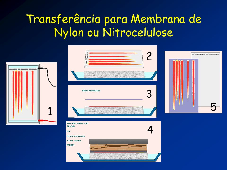
Transferência para Membrana de Nylon ou Nitrocelulose
2 3 5 1 4
24
Hibridização em Nylon ou Nitrocelulose

25
Interpretação de resultados
1 2 3 4 5 6 1 2 3 4 5 6 Sítio alvo para sonda Sítio de restrição Inserção Fonte: IPGRI

26
Herança de RFLPs A B F1 F2 8 Kb 6 Kb 8 Kb 6 Kb 8 Kb 8 Kb 8 Kb 6 Kb

27
RFLP em Cana de Açúcar Fonte: CEBMEG 1 5 10 15 20
This slide shows RFLP marker segregation in Brassica (courtesy of D. Marshall). Each track on the gel was loaded with a separate DNA restriction digest sample. The black bands represent where the radioactive probe hybridised to DNA fragments on the nitrocellulose filter. The difference in banding patterns between the samples reflects genetic differences between the samples. Fonte: CEBMEG
. Each track on the gel was loaded with a separate DNA restriction digest sample. The black bands represent where the radioactive probe hybridised to DNA fragments on the nitrocellulose filter. The difference in banding patterns between the samples reflects genetic differences between the samples. Fonte: CEBMEG.")
28
Vantagens e Desvantagens de RFLP
Reprodutível Marcadores co-dominantes Simples Trabalhoso Caro Uso de sondas radioativas* Fonte: IPGRI

29
Random Amplification of Polymorphic DNA - RAPD
Amplifica seqüências anônimas de DNA usando primers arbitrários 10 bases com >50% G+C PCR com um único primer Método rápido para detecção de polimorfismos Marcador dominante Problemas de reprodutibilidade The random amplified polymorphic DNA (RAPD) technique is a PCR based method which uses one or sometimes two short arbitrary primers (usually 8-10 bases) to amplify anonymous stretches of DNA which are then separated and visualised by gel electrophoresis. The key point about this technique is that nothing is known about the identity of the amplification products. The amplification products are however extremely useful as markers in genetic diversity studies. Other important features of the technique are: The number of fragments. Many different fragments are normally amplified using each single primer, and the technique has therefore proved a fast method for detecting polymorphisms. The majority of commercially produced primers result in 6 to 12 fragments; some primers may fail to give any amplification fragments from some material. Simplicity of the technique. RAPD analysis does not involve hybridisation/autoradiography or high technical expertise. Only tiny quantities of target DNA are required. Arbitrary primers can be purchased. Unit costs per assay are low. This has made RAPD analysis very popular. RAPD markers are dominant. Amplification either occurs at a locus or it does not, leading to scores of band presence/absence; this means that homozygotes and heterozygotes cannot be distinguished. Problems of reproducibility - RAPD does suffer from a sensitivity to changes in PCR conditions resulting in changes to some of the amplified fragments. Reproducible results can be obtained if care is taken to standardise the conditions used (Munthali et al., 1992; Lowe et al., 1996).
 technique is a PCR based method which uses one or sometimes two short arbitrary primers (usually 8-10 bases) to amplify anonymous stretches of DNA which are then separated and visualised by gel electrophoresis. The key point about this technique is that nothing is known about the identity of the amplification products. The amplification products are however extremely useful as markers in genetic diversity studies. Other important features of the technique are: The number of fragments. Many different fragments are normally amplified using each single primer, and the technique has therefore proved a fast method for detecting polymorphisms. The majority of commercially produced primers result in 6 to 12 fragments; some primers may fail to give any amplification fragments from some material. Simplicity of the technique. RAPD analysis does not involve hybridisation/autoradiography or high technical expertise. Only tiny quantities of target DNA are required. Arbitrary primers can be purchased. Unit costs per assay are low. This has made RAPD analysis very popular. RAPD markers are dominant. Amplification either occurs at a locus or it does not, leading to scores of band presence/absence; this means that homozygotes and heterozygotes cannot be distinguished. Problems of reproducibility - RAPD does suffer from a sensitivity to changes in PCR conditions resulting in changes to some of the amplified fragments. Reproducible results can be obtained if care is taken to standardise the conditions used (Munthali et al., 1992; Lowe et al., 1996).")
30
RAPD AA AA Aa aa Aa The random amplified polymorphic DNA (RAPD) technique is a PCR based method which uses one or sometimes two short arbitrary primers (usually 8-10 bases) to amplify anonymous stretches of DNA which are then separated and visualised by gel electrophoresis. The key point about this technique is that nothing is known about the identity of the amplification products. The amplification products are however extremely useful as markers in genetic diversity studies. Other important features of the technique are: The number of fragments. Many different fragments are normally amplified using each single primer, and the technique has therefore proved a fast method for detecting polymorphisms. The majority of commercially produced primers result in 6 to 12 fragments; some primers may fail to give any amplification fragments from some material. Simplicity of the technique. RAPD analysis does not involve hybridisation/autoradiography or high technical expertise. Only tiny quantities of target DNA are required. Arbitrary primers can be purchased. Unit costs per assay are low. This has made RAPD analysis very popular. RAPD markers are dominant. Amplification either occurs at a locus or it does not, leading to scores of band presence/absence; this means that homozygotes and heterozygotes cannot be distinguished. Problems of reproducibility - RAPD does suffer from a sensitivity to changes in PCR conditions resulting in changes to some of the amplified fragments. Reproducible results can be obtained if care is taken to standardise the conditions used (Munthali et al., 1992; Lowe et al., 1996). aa
 technique is a PCR based method which uses one or sometimes two short arbitrary primers (usually 8-10 bases) to amplify anonymous stretches of DNA which are then separated and visualised by gel electrophoresis. The key point about this technique is that nothing is known about the identity of the amplification products. The amplification products are however extremely useful as markers in genetic diversity studies. Other important features of the technique are: The number of fragments. Many different fragments are normally amplified using each single primer, and the technique has therefore proved a fast method for detecting polymorphisms. The majority of commercially produced primers result in 6 to 12 fragments; some primers may fail to give any amplification fragments from some material. Simplicity of the technique. RAPD analysis does not involve hybridisation/autoradiography or high technical expertise. Only tiny quantities of target DNA are required. Arbitrary primers can be purchased. Unit costs per assay are low. This has made RAPD analysis very popular. RAPD markers are dominant. Amplification either occurs at a locus or it does not, leading to scores of band presence/absence; this means that homozygotes and heterozygotes cannot be distinguished. Problems of reproducibility - RAPD does suffer from a sensitivity to changes in PCR conditions resulting in changes to some of the amplified fragments. Reproducible results can be obtained if care is taken to standardise the conditions used (Munthali et al., 1992; Lowe et al., 1996). aa.")
31
Interpretação de RAPDs
Marcadores RAPD são anônimos Dados binários (presença x ausência) RAPD são dominantes (AA = Aa) Problemas de co-migração mesma banda, mesmo fragmento? uma banda, um fragmento?
 RAPD são dominantes (AA = Aa) Problemas de co-migração. mesma banda, mesmo fragmento uma banda, um fragmento")
32
RAPD - resumo Rápido Simples Baixo custo Sem uso de radioisótopos
Marcador dominante Problemas de reprodutibilidade Problemas de interpretação Fonte: IPGRI

33
RAPD - primer OPJ04 - Bananeira
1500 pb

34
RAPD - Feijoeiro OPY04 OPAM13

Apresentações semelhantes


 dos genes e seu armazenamento>")


>")


