Carregar apresentação
A apresentação está carregando. Por favor, espere

1
REUNIÃO ANATOMOENDOSCÓPICA
SERVIÇO DE ENDOSCOPIA GASTROINTESTINAL HOSPITAL DAS CLÍNICAS FACULDADE DE MEDICINA DA UNIVERSIDADE DE SÃO PAULO REUNIÃO ANATOMOENDOSCÓPICA E1 VINICIUS LEITE DE CASTRO DR. KENDI YAMAZAKI DRA. ELISA RYOKA BABA SÃO PAULO MARÇO 2013

2
CASO CLÍNICO M. P. G. ; 64 a ; feminino
Dor abdominal difusa iniciada há 1 ano do tipo cólica, sem irradiação, de moderada intensidade associada à constipação intestinal, inapetência e perda ponderal de 5 kg nos últimos 6 meses Colecistectomia convencional; Cesariana Tabagista 40 anos-maço

3
EXAME FÍSICO Descorada (+/4+) Ausculta pulmonar sem alterações
Abdome doloroso à palpação em epigástrio, hipocôndrio e flanco esquerdos Ausência de visceromegalias ou massas palpáveis

4
CT ABDOMINAL

5
CT ABDOMINAL

6
CT ABDOMINAL Infiltração e espessamento parietal, reduzindo luz do cólon no ângulo esplênico, medindo 7,5 cm de extensão, compatível com neoplasia primária. Linfonodomegalias mesentéricas, destacando-se conglomerado medindo 2,8 x 2,2 cm em contato com grande curvatura gástrica. Fígado com lesões hipoatenuantes no segmento VII medindo 0,8 cm e IV com 0,5 cm, incaracterísticas. Veia porta pérvia. Raros divertículos no cólon descendente / sigmóide

7
CT TORÁCICA Raras bolhas de enfisema nos ápices pulmonares

8
ENDOSCOPIA DIGESTIVA ALTA

9
ENDOSCOPIA DIGESTIVA ALTA
Estômago: Pólipo séssil em fundo gástrico, coberto por mucosa normal de 5 mm. Polipectomia. Em corpo distal, para a grande curvatura, nota-se área de enantema e edema da mucosa. Biópsias. Anatomopatológico: Pólipo: EROSÃO REPARADA Área de enantema e edema: GASTRITE CRÔNICA LEVE, COM EDEMA DE LÂMINA PRÓPRIA, EM MUCOSA DE PADRÃO FÚNDICO Helicobacter pylori: NEGATIVO

10
COLONOSCOPIA

11
COLONOSCOPIA Progressão até ângulo esplênico, observando-se lesão ulceroinfiltrativa com áreas de tecido necrótico, circunferencial, impedindo a progressão do aparelho. Biópsias. Descendente e sigmóide com múltiplos óstios diverticulares, sem sinais inflamatórios e/ou sangramento Reto distal observa-se lesão elevada, subepitelial, amarelada e endurecida, de 8 mm, a 8 cm da borda anal. Biópsias.

12
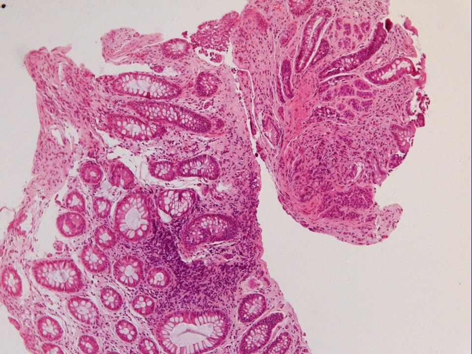
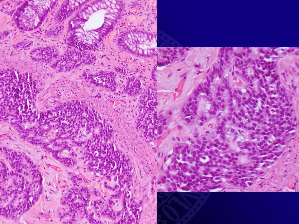
ANATOMOPATOLÓGICO

13
ANATOMOPATOLÓGICO

15
CDX2 CK20 CK7 cromogranina

16
Ki67

17
ANATOMOPATOLÓGICO Lesão de ângulo esplênico: ADENOCARCINOMA TUBULO-VILOSO MODERADAMENTE DIFERENCIADO, ULCERADO Lesão de reto: TUMOR NEUROENDÓCRINO BEM DIFERENCIADO (GRAU 1) Imunohistoquímica CDX2 ; Citoqueratina 20 ; Citoqueratina 7 ; Cromogranina +; Ki67 + (< 2%) Perfil compatível com tumor neuroendócrino bem diferenciado Grau 1
 Imunohistoquímica. CDX2 ; Citoqueratina 20 ; Citoqueratina 7 ; Cromogranina +; Ki67 + (< 2%) Perfil compatível com tumor neuroendócrino bem diferenciado Grau 1.")
18
COLONOSCOPIA

19
COLONOSCOPIA Realizada mucosectomia seguida de tatuagem do leito de ressecção com tinta nanquim.

20
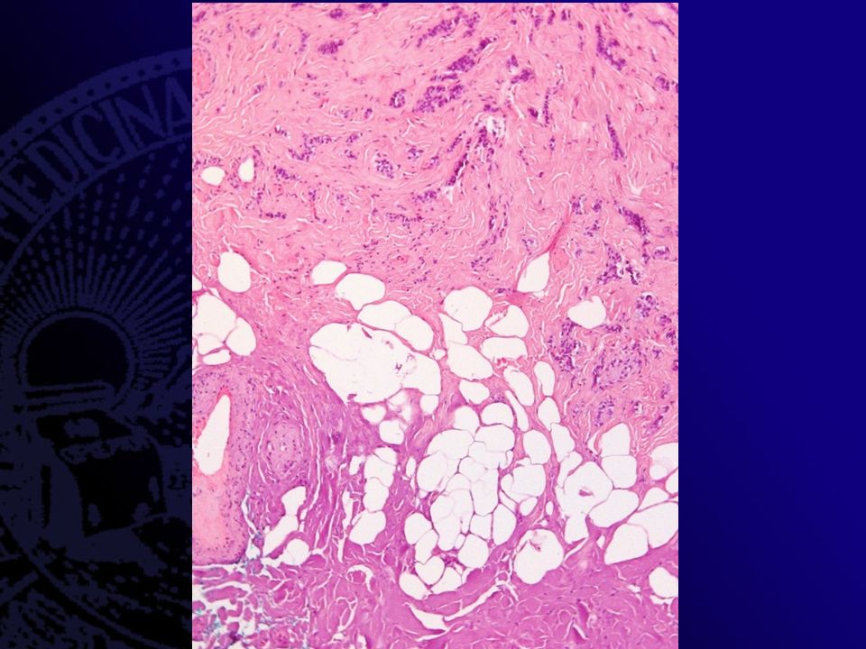
ANATOMOPATOLÓGICO

24
Ki67

25
ANATOMOPATOLÓGICO Tumor neuroendócrino bem diferenciado, medindo 0,4 cm no maior eixo Margens cirúrgicas livres de comprometimento neoplásico Imunohistoquímica Ki-67 + (<2%), Cromogranina +, Sinaptofisina + Perfil compatível com tumor neuroendócrino bem diferenciado Grau 1
, Cromogranina +, Sinaptofisina + Perfil compatível com tumor neuroendócrino bem diferenciado Grau 1.")
26
EVOLUÇÃO 27/02/2013: Colectomia esquerda + esplenectomia + biópsia hepática + lavado peritoneal Anatomopatológico ADENOCARCINOMA TUBULAR BEM DIFERENCIADO, medindo 5,0 x 3,7 x 1,3 cm, infiltração até subserosa, sem invasão angiolinfática. Margens de ressecção cirúrgica proximal e distal livres (9 cm e 2 cm, respectivamente). Ausência de metástase em 14 linfonodos examinados. Estadiamento (TNM): pT3, pN0
. Ausência de metástase em 14 linfonodos examinados. Estadiamento (TNM): pT3, pN0.")
27
EVOLUÇÃO POI em UTI 5º PO = deiscência da anastomose
Laparotomia exploradora: colectomia segmentar + colostomia (transverso) + amputação de coto retal (Hartmann) Anatomopatológico: Segmentos colônicos exibindo processo inflamatório crônico inespecífico com área de ulceração da mucosa em anastomose; serosite aguda fibrino purulenta com esteatonecrose; linfonodos com hiperplasia linfóide reativa 11º PO eletiva / 6º PO urgência Alta Hospitalar
 + amputação de coto retal (Hartmann) Anatomopatológico: Segmentos colônicos exibindo processo inflamatório crônico inespecífico com área de ulceração da mucosa em anastomose; serosite aguda fibrino purulenta com esteatonecrose; linfonodos com hiperplasia linfóide reativa. 11º PO eletiva / 6º PO urgência Alta Hospitalar.")
28
TUMOR NEUROENDÓCRINO RETAL

29
INTRODUÇÃO Sistema neuroendócrino Todo sistema gastroenteropancreático
Maior órgão endócrino tipos celulares / 40 tipos de hormônios ou aminas bioativas Sistema nervoso autônomo Neoplasias: tumor carcinóide ou células das ilhotas Neoplasia ou tumor neuroendócrino (NET) Carcinoma neuroendócrino (NEC) Jernman J.; Välimäki M.J.; Louhimo J; et al. The Novel WHO 2010 Classification for Gastrointestinal Neuroendocrine Tumours Correlates Well with the Metastatic Potential of Rectal Neuroendocrine Tumours. Neuroendocrinology 2012;95:317–324 Sistema neuroendócrino encontra-se difuso no aparelho digestivo; em conjunto as células constituem o maior órgão endócrino do organismo, constituído por cerca de 19 tipos celulares e capaz de produzir pelo menos 40 tipos de substâncias que incluem hormônios ou aminas ativas SOB estímulo do sistema nervoso autônomo e assim regulando o sistema gastrointestinal; Neoplasms that arise from these cells used to be called carcinoid tumours; the appropriate modern term is neuroendocrine neoplasm, neuroendocrine tumour (NET) or neuroendocrine carcinoma (NEC).
 Carcinoma neuroendócrino (NEC) Jernman J.; Välimäki M.J.; Louhimo J; et al. The Novel WHO 2010 Classification for Gastrointestinal Neuroendocrine Tumours Correlates Well with the Metastatic Potential of Rectal Neuroendocrine Tumours. Neuroendocrinology 2012;95:317–324. Sistema neuroendócrino encontra-se difuso no aparelho digestivo; em conjunto as células constituem o maior órgão endócrino do organismo, constituído por cerca de 19 tipos celulares e capaz de produzir pelo menos 40 tipos de substâncias que incluem hormônios ou aminas ativas SOB estímulo do sistema nervoso autônomo e assim regulando o sistema gastrointestinal; Neoplasms that arise from these cells used to be called carcinoid tumours; the appropriate modern term is neuroendocrine neoplasm, neuroendocrine tumour (NET) or neuroendocrine carcinoma (NEC).")
30
PRODUTOS SECRETADOS PELO TUMOR CARCINÓIDE (TUMOR NEUROENDÓCRINO)
INTRODUÇÃO PRODUTOS SECRETADOS PELO TUMOR CARCINÓIDE (TUMOR NEUROENDÓCRINO) AMINAS BIOATIVAS Serotonina (5-hidroxitriptamina) Histamina Dopamina Norepinefrina PEPTÍDIOS ACTH; GHRH Calcitonina Polipeptídio pancreático Bradicinina Neurotensina Cromogranina Secretina Colecistocinina Calicreína Gastrina Insulina Prostaglandinas Endoderma Pulmão e ovário Expressão celular específica Hormônios Marcadores gerais Sinaptofisina Cromogranina A Não funcionante Embriogênse ENDODERMA podemos encontrar no pulmão e ovário Caracterizados pela expressão de de células específicas do tipo de hormônios péptidicos e marcadores gerais (sinaptofisina, cromogranina A)
 AMINAS BIOATIVAS. Serotonina (5-hidroxitriptamina) Histamina. Dopamina. Norepinefrina. PEPTÍDIOS. ACTH; GHRH. Calcitonina. Polipeptídio pancreático. Bradicinina. Neurotensina. Cromogranina. Secretina. Colecistocinina. Calicreína. Gastrina. Insulina. Prostaglandinas. Endoderma. Pulmão e ovário. Expressão celular específica. Hormônios. Marcadores gerais. Sinaptofisina. Cromogranina A. Não funcionante. Embriogênse ENDODERMA podemos encontrar no pulmão e ovário. Caracterizados pela expressão de de células específicas do tipo de hormônios péptidicos e marcadores gerais (sinaptofisina, cromogranina A)")
31
FATORES DE RISCO Álcool e tabagismo Sexo feminino e história familiar
Chen CC, Neugut AI, Rotterdam H. Risk factors for adenocarcinomas and malignant carcinoids of the small instestine: preliminary findings. Cancer Epidemiol Biomarkers Prev 1994;3:205-7 Sexo feminino e história familiar Hassan MM, Phan A, Li D et al. Risk factors associated with neuroendocrine tumors: A U.S.-based case-control study. Int J Cancer 2008;123:867-73

32
NET RETAL Pequenos (<1 cm) Não funcionais
Móveis em relação à musculatura subjacente Malignidade Crescimento lento x agressividade Yao JC, Hassan M, Phan A, et al.: One hundred years after ‘carcinoid’: epidemiology of and prognostic factors for neuroendocrine tumors in 35,825 cases in the United States. J Clin Oncol 2008; 26: 3063–3072. São geralmente pequenos (<1 cm), não funcionais, móveis em relação à musculatura subjacente e podem ser ressecados endoscopicamente Os tumores carcinoides gastrointestinais são malignos, em geral de crescimento lento quando comparados ao adenocarcinoma mas podem comportar-se de maneira agressiva
, não funcionais, móveis em relação à musculatura subjacente e podem ser ressecados endoscopicamente. Os tumores carcinoides gastrointestinais são malignos, em geral de crescimento lento quando comparados ao adenocarcinoma mas podem comportar-se de maneira agressiva.")
33
NET RETAL Incidência anual de 0,83 / 100.000 habitantes nos EUA
Yao JC, Hassan M, Phan A, et al.: One hundred years after ‘carcinoid’: epidemiology of and prognostic factors for neuroendocrine tumors in 35,825 cases in the United States. J Clin Oncol 2008; 26: 3063–3072. 13,7% dos NET Modlin IM, Lye KD, Kidd M: A 5-decade analysis of 13,715 carcinoid tumors. Cancer 2003; 97: 934–959 Retal tumors have an age-adjusted annual incidence of 0.83 per 100,000 in the US

34
IMUNOHISTOQUÍMICA 80% expressam peptídio pancreático (PP)
30% positivos para serotonina Fiocca R, Rindi G, Capella C, Grimelius L, Polak JM, Schwartz TW, Yanaihara N, Solcia E: Glucagon, glicentin, proglucagon, PYY, PP and proPP-icosapeptide immunoreactivities of rectal carcinoid tumors and related non-tumor cells. Regul Pept 1987; 17: 9–29. ENETS¹ imunorreatividade para cromogranina A e sinaptofisina Klöppel et al.² Soga et al.³ 60.9% positivos para cromogranina A Enetes ( European Neuroendocrine Tumor Society) x Kloppel x Soga The ENETS recommends verificação imunohistoquímica da natureza neuroendócrina com chromogranin A and synaptophysin Klöppel et al. stated that NETs of the rectum are immunoreactive for synaptophysin but negative for chromogranin Soga reported that 60.9% of rectal carcinoids were positive for chromogranin A.
 x Kloppel x Soga. The ENETS recommends verificação imunohistoquímica da natureza neuroendócrina com chromogranin A and synaptophysin. Klöppel et al. stated that NETs of the rectum are immunoreactive for synaptophysin but negative for chromogranin. Soga reported that 60.9% of rectal carcinoids were positive for chromogranin A.")
35
RASTREAMENTO Aumento na detecção
Endoscopia e imagens de alta resolução Imunohistoquímica > 50% Incidental / Estadio precoce Scherubl H. Rectal carcinoids are on the rise: early detection by screening endoscopy. Endoscopy 2009;41:162–165 Doença local Sobrevida em 5 anos de % Yao JC, Hassan M, Phan A, et al.: One hundred years after ‘carcinoid’: epidemiology of and prognostic factors for neuroendocrine tumors in 35,825 cases in the United States. J Clin Oncol 2008; 26: 3063–3072. The detection of rectal carcinoids has increased with the greater awareness by clinicians and pathologists (imunohistoquímica) and with the more sophisticated diagnostic tools, and more than half of all carcinoid tumors in the rectum are incidentally diagnosed at an early stage Rectal NETs are usually small and are often discovered by chance during endoscopy. Patients with local disease have a favourable prognosis with a 5-year survival rate of 88–91%
 and with the more sophisticated diagnostic tools, and more than half of all carcinoid tumors in the rectum are incidentally diagnosed at an early stage. Rectal NETs are usually small and are often discovered by chance during endoscopy. Patients with local disease have a favourable prognosis with a 5-year survival rate of 88–91%")
36
Screening sigmoidoscopy and colonoscopy are highly effective not only in the early detection of colorectal adenomas and adenocarcinomas but also in the early detection of neuroendocrine neoplasms of the (colo−)rectum. This is in line with the steadily improving overall 5−year survival observed by Modlin and coworkers Modlin IM, Lye KD, Kidd M. A 5-decade analysis of 13,715 carcinoid tumors. Cancer 2003;97:934–959.

37
QUADRO CLÍNICO Obstrução Sangramento Perfuração Os sintomas dependem:
atividade hormonal localização extensão efeito de massa, sangramento, obstrução ou até perfuração Suyama K. et al. Neuroendocrine tumor of the rectum. The American Journal of Surgery 2009; 198: e39-e41

38
CLASSIFICAÇÃO WHO 2010 x WHO 2000 potencial metastático e prognóstico Jernman J.; Välimäki M.J.; Louhimo J; et al. The Novel WHO 2010 Classification for Gastrointestinal Neuroendocrine Tumours Correlates Well with the Metastatic Potential of Rectal Neuroendocrine Tumours. Neuroendocrinology 2012;95:317–324 Carcinóide retal Tumor neuroendócrino ou carcinoma neuroendócrino? Scherübl H. Rectal carcinoids are on the rise: early detection by screening endoscopy. Endoscopy 2009; 41: Em nosso trabalho envolvendo 73 NET, a classificação de 2010 mostrou-se superior a de 2000 em predizer o potencial metastático e estabelecer o prognóstico. NENs Bem diferenciados são classificados juntos como tumores neuroendócrinos (TNE) G1 ou G2 O termo carcinoma neuroendócrino (CNE), refere-se a todos as NENs pouco diferenciados CNE subdividido (pequenas-células e grandes-células) As concerns nomenclature, (well−differentiated) neuroendocrine neoplasms of the rectum that either show angioinvasion or infiltration of the muscularis propria (or beyond) or have metastasized are called neuroendocrine carcinomas. The term “rectal carcinoid” does not distinguish between well−differentiated neuroendocrine carcinoma and well−differentiated neuroendocrine tumor (of the rectum); it comprises both
 G1 ou G2. O termo carcinoma neuroendócrino (CNE), refere-se a todos as NENs pouco diferenciados. CNE subdividido (pequenas-células e grandes-células) As concerns nomenclature, (well−differentiated) neuroendocrine neoplasms of the rectum that either show angioinvasion or infiltration of the muscularis propria (or beyond) or have metastasized are called neuroendocrine carcinomas. The term rectal carcinoid does not distinguish between well−differentiated neuroendocrine carcinoma and well−differentiated neuroendocrine tumor (of the rectum); it comprises both.")
39
CLASSIFICAÇÃO Grau 1 e Grau 2 Neoplasia ou tumor neuroendócrino
In the novel WHO 2010 classification for GI NETs, all tumours are considered malignant with the potential to metastasize. In this classification, tumour GRADE IS BASED ON THE PROLIFERATION INDEX AND MITOTIC COUNT. Divided into 3 groups. Grau 1 e Grau 2 Bem diferenciados. Grau 3 Pouco diferenciados Carcinoma Neuroendócrino (dividios em pequenas-células e grande-células) Jernman J.; Välimäki M.J.; Louhimo J; et al. The Novel WHO 2010 Classification for Gastrointestinal Neuroendocrine Tumours Correlates Well with the Metastatic Potential of Rectal Neuroendocrine Tumours. Neuroendocrinology 2012;95:317–324 Grau 1 e Grau 2 Neoplasia ou tumor neuroendócrino Grau 3 Carcinoma Neuroendócrino Pequenas-células x Grandes-células
 Jernman J.; Välimäki M.J.; Louhimo J; et al. The Novel WHO 2010 Classification for Gastrointestinal Neuroendocrine Tumours Correlates Well with the Metastatic Potential of Rectal Neuroendocrine Tumours. Neuroendocrinology 2012;95:317–324. Grau 1 e Grau 2 Neoplasia ou tumor neuroendócrino. Grau 3 Carcinoma Neuroendócrino. Pequenas-células x Grandes-células.")
40
ESTADIAMENTO Endoscopia
Cintilografia com receptores de somatostatina (octreoscan) CT helicoidal ou MRI Scherübl H. Rectal carcinoids are on the rise: early detection by screening endoscopy. Endoscopy 2009; 41: Ecoendoscopia Diferenciação lesões subepiteliais Monitoramento dos tumores Thomas-Marques, L., et al., Prospective endoscopic ultrasonographic evaluation of the frequency of nonfunctioning pancreaticoduodenal endocrine tumors in patients with multiple endocrine neoplasia type 1. Am J Gastroenterol, (2): p In light of the risk of metastases, nowadays we SHOULD stage all patients with carcinoids before deciding on treatment. Importância da ecoendoscopia na detecção precoce (diferenciação com outras lesões subepiteleiais ) e monitoramento desses tumores, em especial nos não-funcionantes (no caso gastrointestinal)
 CT helicoidal ou MRI. Scherübl H. Rectal carcinoids are on the rise: early detection by screening endoscopy. Endoscopy 2009; 41: Ecoendoscopia. Diferenciação lesões subepiteliais. Monitoramento dos tumores. Thomas-Marques, L., et al., Prospective endoscopic ultrasonographic evaluation of the frequency of nonfunctioning pancreaticoduodenal endocrine tumors in patients with multiple endocrine neoplasia type 1. Am J Gastroenterol, (2): p In light of the risk of metastases, nowadays we SHOULD stage all patients with carcinoids before deciding on treatment. Importância da ecoendoscopia na detecção precoce (diferenciação com outras lesões subepiteleiais ) e monitoramento desses tumores, em especial nos não-funcionantes (no caso gastrointestinal)")
41
METÁSTASE Linfonodos regionais; fígado; pulmão; osso; peritônio
Taxas similares entre carcinomas GI e NETs GI Soga J. Early-stage carcinoids of the gastrointestinal tract: an analysis of 1914 reported cases. Cancer 2005;103:1587–1595 SEER 9% metástase ao diagnóstico Yao JC, Hassan M, Phan A, et al.: One hundred years after ‘carcinoid’: epidemiology of and prognostic factors for neuroendocrine tumors in 35,825 cases in the United States. J Clin Oncol 2008; 26: 3063–3072. The Surveillance, Epidemiology, and End Results (SEER) registry database of the National Cancer Institute (which reflects the standard of care for the average US citizen) – O que significa o SEER Metastasis rates were similar for gastrointestinal carcinomas and gastrointestinal carcinoids According to the SEER (Surveillance, Epidemiology and End Results) database, 9% of the patients with a rectal NET have disseminated disease
 registry database of the National Cancer Institute (which reflects the standard of care for the average US citizen) – O que significa o SEER. Metastasis rates were similar for gastrointestinal carcinomas and gastrointestinal carcinoids. According to the SEER (Surveillance, Epidemiology and End Results) database, 9% of the patients with a rectal NET have disseminated disease.")
42
METÁSTASE ≤ 10 mm 3% - 9,8% 10,1 – 20 mm 17% - 81% para linfonodos regionais > 20 mm 60% - 80% Scherübl H. Rectal carcinoids are on the rise: early detection by screening endoscopy. Endoscopy 2009; 41: Modlin I, Lye K, Kidd M. A 5−decade analysis of carcinoid tumors. Cancer 2003; 97: 1) 60%±80% of rectal carcinoids that are larger than 2 cm metastasize 2) rectal carcinoids that are 10.1±20mm in diameter spread to regional lymph nodes in 17%±81% of patients 3) rectal carcinoids that are 1 cm or less in size metastasize in 3%±9.8% of cases
 60%±80% of rectal carcinoids that are larger than 2 cm metastasize 2) rectal carcinoids that are 10.1±20mm in diameter spread to regional lymph nodes in 17%±81% of patients 3) rectal carcinoids that are 1 cm or less in size metastasize in 3%±9.8% of cases.")
43
TRATAMENTO Ressecção endoscópica
Ishii et al. e Yamaguchi et al. ≤ 10 mm Park et al. < 16 mm Kwaan et al. < 20 mm Ishii N, Horiki N, Itoh T et al. Endoscopic submucosal dissection and preoperative assessment with endoscopic ultrasonography for the treatment of rectal carcinoid tumors. Surg Endosc 2010;24:1413–1419. Yamaguchi N, Isomoto H, Nishiyama et al. Endoscopic submucosal dissection for rectal carcinoid tumors. Surg Endosc 2010;24:504–508. Park HW, Byeon JS, Park YS, et al. Endoscopic submucosal dissection for treatment of rectal carcinoid tumors. Gastrointest Endosc 2010;72:143–149. Kwaan MR, Goldberg JE, Bleday R. Rectal carcinoid tumors: review of results after endoscopic and surgical therapy. Arch Surg 2008;143:471–475. Previous reports have presented different size indications for rectal carcinoid endoscopic resection: ≤ 10mm (Ishii et al. and Yamaguchi et al.), < 16mm (Park et al.), and < 20mm (Kwaan et al.).
, < 16mm (Park et al.), and < 20mm (Kwaan et al.).")
44
TRATAMENTO Ecoendoscopia tamanho exato e grau de invasão
Sem ecoendoscopia ou apenas técnicas endoscópicas convencionais Margens de ressecção indeterminadas ou positivas em 31,8% - 83% dos pacientes Kwaan M, Goldberg J, Bleday R et al. Rectal carcinoid tumors: review of results after endoscopic and surgical therapy. Arch Surg 2008; 143: Kim YJ, Lee SK, Cheon JH et al. Efficacy of endoscopic resection for small rectal carcinoid: a retrospective study. Korean J Gastroenterol 2008; 51: Ecoendoscopia previamente à ressecção taxas de 4,8% - 17% Mashimo Y, Matsuda T, Uraoka T et al. Endoscopic submucosal resection with a ligation device is an effective and safe treatment for carcinoid tumors in the lower rectum. J Gastroenterol Hepatol 2008; 23: Kobayashi K, Katsumata T, Yoshizawa S et al. Indications of endoscopic polypectomy for rectal carcinoid tumors and clinical usefulness of endoscopic ultrasonography. Dis Colon Rectum 2005; 48: 285 – 291 Before embarking on resection, the exact tumor size and in particular the depth of invasion has to be determined by endoscopic ultrasound When either endoscopic ultrasonography was omitted in tumor staging or conventional endoscopic techniques were used, indeterminate or even positive resection margins were observed on histological examination in as many as 31.8%±83% of patients When endoscopic ultrasonography was performed prior to endoscopic submucosal resection, the R1 rate dropped to 4.8%±17%

45
TRATAMENTO T1aN0 (≤ 1 cm; sem invasão submucosa) excisão local endoscópica ou cirúrgica Okamoto Y, Fujii M, Tateiwa S, et al. Treatment of multiple rectal carcinoids by endoscopic mucosal resection using a device for esophageal variceal ligation. Endoscopy 2004;36:469–470. Moon, SH. et al. Endoscopic Submucosal dissection for rectal neuroendocrine (carcinoid) tumors. Jounal of Laparoendoscopic & advanced surgical techniques. 2011; 21(8): 695 – 699. ESD x EMR Lee DS., et al. The feasibility of endoscopic submucosal dissection for rectal carcinoid tumors: comparison with endoscopic mucosal resection. Endoscopy 2010; 42: 647 – 651 Onozato Y., Kakizaki S., Ishihara H., et al. Endoscopic submucosal dissection for rectal tumors. Endoscopy 2007; 39: 423 – 427 Previous studies have shown that T1aN0 rectal carcinoid tumors < 1 cm in size and located within the submucosa without metastasis can be managed using local endoscopic or transanal resection. excisão local endoscópica ou cirúrgica Superioridade da ESD sobre a EMR para tratmento local de tumores carcinoides retais < 10 mm
 tumors. Jounal of Laparoendoscopic & advanced surgical techniques. 2011; 21(8): 695 – 699. ESD x EMR. Lee DS., et al. The feasibility of endoscopic submucosal dissection for rectal carcinoid tumors: comparison with endoscopic mucosal resection. Endoscopy 2010; 42: 647 – 651. Onozato Y., Kakizaki S., Ishihara H., et al. Endoscopic submucosal dissection for rectal tumors. Endoscopy 2007; 39: 423 – 427. Previous studies have shown that T1aN0 rectal carcinoid tumors < 1 cm in size and located within the submucosa without metastasis can be managed using local endoscopic or transanal resection. excisão local endoscópica ou cirúrgica. Superioridade da ESD sobre a EMR para tratmento local de tumores carcinoides retais < 10 mm.")
46
ESD Endoscopic submucosal dissection of a rectal carcinoid tumor. (A) An 8-mm rectal carcinoid tumor. (B) A solution of 10% glycerin and 5% fructose in 0.9% saline was injected to lift the tumor. (C, D) Mucosal incision and submucosal dissection. (E) A clear ulcer after complete tumor removal. (F) Macroscopic view of the resected specimen (13 · 12 mm). Moon, SH. et al. Endoscopic Submucosal dissection for rectal neuroendocrine (carcinoid) tumors. Jounal of Laparoendoscopic & advanced surgical techniques. 2011; 21(8): 695 – 699.
 Mucosal incision and submucosal dissection. (E) A clear ulcer after complete tumor removal. (F) Macroscopic view of the resected specimen (13 · 12 mm). Moon, SH. et al. Endoscopic Submucosal dissection for rectal neuroendocrine (carcinoid) tumors. Jounal of Laparoendoscopic & advanced surgical techniques. 2011; 21(8): 695 – 699.")
47
TRATAMENTO T1bN0 (10,1 – 20 mm) sem consenso
Kwaan MR, Goldberg JE, Bleday R. Rectal carcinoid tumors: review of results after endoscopic and surgical therapy. Arch Surg 2008;143:471–475. Japão ressecção cirúrgica similar aos adenocarcinomas Linfadenectomia regional tratamento padrão Konishi T, Watanabe T, Kishimoto J et al. Prognosis and risk factors of metastasis in colorectal carcinoids: results of a nationwide registry over 15 years. Gut. 2007; 56: Konishi T, Watanabe T, Kishimoto J et al. Prognosis and metastatic potential of colorectal carcinoids compared with adenocarcinomas: results of a nationwide registry over 15 years. J Clin Oncol 2008; 26: ASCO Abstract 4054 Tsukamoto S, Fujita S, Yamaguchi T et al. Clinicopathological characteristics and prognosis of rectal well−differentiated neuroendocrine tumors. Int J Colorectal Dis 2008; 23: For rectal carcinoids of T1bN0 (the same as T1aN0 except sized between 1 and 2 cm), there is no consensus in the literature concerning the appropriate therapy therapy of rectal carcinoids measuring 10.1±20mmin diameter is a matter of debate; There are no controlled prospective studies on local teraphy of rectal carcinoids; In Japan rectal carcinoids that are larger than 10.0mm are treated surgically along the same lines as rectal adenocarcinomas . There, lymph node dissection is considered the standard of care for rectal carcinoids 10.1±20.0mm in size. Seguem princípios oncológicos de ressecção
, there is no consensus in the literature concerning the appropriate therapy. therapy of rectal carcinoids measuring 10.1±20mmin diameter is a matter of debate; There are no controlled prospective studies on local teraphy of rectal carcinoids; In Japan rectal carcinoids that are larger than 10.0mm are treated surgically along the same lines as rectal adenocarcinomas . There, lymph node dissection is considered the standard of care for rectal carcinoids 10.1±20.0mm in size. Seguem princípios oncológicos de ressecção.")
48
TRATAMENTO T2 (> 20 mm) ou ulcerados Ressecção cirúrgica com linfadenectomia Metástase cirurgias citorredutoras hepáticas QT (estreptozotocina e 5-fluorouracil) Quimioembolização Rectal carcinoids larger than 2 cm and Ulcerated colorectal carcinoids CIRURGIA COM LINFADENECTOMIA Seguem princípios oncológicos de ressecção Nos casos de metástase, cirurgias citorredutoras hepáticas podem melhorar índice de sucesso de tratamento subsequente: quimioterapia (estreptozotocina e 5-fluorouracil); quimioembolização;
 Quimioembolização. Rectal carcinoids larger than 2 cm and Ulcerated colorectal carcinoids CIRURGIA COM LINFADENECTOMIA. Seguem princípios oncológicos de ressecção. Nos casos de metástase, cirurgias citorredutoras hepáticas podem melhorar índice de sucesso de tratamento subsequente: quimioterapia (estreptozotocina e 5-fluorouracil); quimioembolização;")
49
SOBREVIDA Sobrevida em 5 anos
Linfonodo negativo, sem invasão vascular ou infiltração de submucosa 98,9% - 100% Linfonodo positivo 54% - 73% Metástase 15% - 30% Modlin I, Drozdov I, Gustafsson B et al. Rectal neuroendocrine tumors - diagnosis and treatment. In: Modlin I, Oberg K (eds). A century of advances in neuroendocrine tumor biology and treatment. Hannover:Felsenstein CCCP, 2007: Soga J. Early−stage carcinoids of the gastrointestinal tract: an analysis of 1914 reported cases. Cancer 2005; 103: Konishi T, Watanabe T, Kishimoto J et al. Prognosis and risk factors of metastasis in colorectal carcinoids: results of a nationwide registry over 15 years. Gut. 2007; 56: The 5−year survival of patients with rectal carcinoid disease who have distant metastases is 15%±30% For node−positive rectal carcinoid disease (without distant metastases at the time of diagnosis), 5−year survival is 54%±73% Histologically node−negative rectal carcinoids that are smaller than 1 cm and do not show angioinvasion or infiltration of the muscularis propria are associated with an excellent 5−year survival of 98.9%±100%
. A century of advances in neuroendocrine tumor biology and treatment. Hannover:Felsenstein CCCP, 2007: Soga J. Early−stage carcinoids of the gastrointestinal tract: an analysis of 1914 reported cases. Cancer 2005; 103: Konishi T, Watanabe T, Kishimoto J et al. Prognosis and risk factors of metastasis in colorectal carcinoids: results of a nationwide registry over 15 years. Gut. 2007; 56: The 5−year survival of patients with rectal carcinoid disease who have distant metastases is 15%±30% For node−positive rectal carcinoid disease (without distant metastases at the time of diagnosis), 5−year survival is 54%±73% Histologically node−negative rectal carcinoids that are smaller than 1 cm and do not show angioinvasion or infiltration of the muscularis propria are associated with an excellent 5−year survival of 98.9%±100%")
50
FOLLOW-UP Seguimento de 10 anos metástase tardia
Kwaan MR, Goldberg JE, Bleday R: Rectal carcinoid tumors: review of results after endoscopic and surgical therapy. Arch Surg 2008; 143: 471–475. G1 follow-up endoscópico simples (excluir recidiva local) G2 Endoscopias repetidas + métodos radiológicos Jernman J.; Välimäki M.J.; Louhimo J; et al. The Novel WHO 2010 Classification for Gastrointestinal Neuroendocrine Tumours Correlates Well with the Metastatic Potential of Rectal Neuroendocrine Tumours. Neuroendocrinology 2012;95:317–324 A follow-up of up to 10 years is recommended, because metastatic lesions may occur late A single follow-up endoscopy to exclude local recurrence seems sufficient in patients with a G1 tumour. Patients with a G2 tumour, on the other hand, need to be monitored much more carefully. Repeated endoscopies alone are insufficient for monitoring these patients, but metastatic lesions should also be excluded using modern imaging techniques. When patients are chosen for intensive long-term follow-up, reliable classification of the primary tumour (including size, dissemination of disease, and proliferation activity) is essential. G1 NETs have low, if any, metastatic potential, whereas G2 NETs often metastasise, so such patients require intensive long-term follow-up.
 G2 Endoscopias repetidas + métodos radiológicos. Jernman J.; Välimäki M.J.; Louhimo J; et al. The Novel WHO 2010 Classification for Gastrointestinal Neuroendocrine Tumours Correlates Well with the Metastatic Potential of Rectal Neuroendocrine Tumours. Neuroendocrinology 2012;95:317–324. A follow-up of up to 10 years is recommended, because metastatic lesions may occur late. A single follow-up endoscopy to exclude local recurrence seems sufficient in patients with a G1 tumour. Patients with a G2 tumour, on the other hand, need to be monitored much more carefully. Repeated endoscopies alone are insufficient for monitoring these patients, but metastatic lesions should also be excluded using modern imaging techniques. When patients are chosen for intensive long-term follow-up, reliable classification of the primary tumour (including size, dissemination of disease, and proliferation activity) is essential. G1 NETs have low, if any, metastatic potential, whereas G2 NETs often metastasise, so such patients require intensive long-term follow-up.")
51
¹ Ramage JK, Goretzki PE, Manfredi R, Komminoth P, Ferone D, Hyrdel R, Kaltsas G, Kelestimur F, Kvols L, Scoazec JY, Garcia MI, Caplin ME, Frascati Consensus Conference participants: Consensus guidelines for the management of patients with digestive neuroendocrine tumours: well-differentiated colon and rectum tumour/carcinoma. Neuroendocrinology 2008; 87: 31–39 ² Klöppel G, Rindi G, Anlauf M, Perren A, Komminoth P: Site-specific biology and pathology of gastroenteropancreatic neuroendocrine tumors. Virchows Arch 2007; 451(suppl 1):S9–S27 ³ Soga J: Carcinoids of the rectum: an evaluation of 1,271 reported cases. Surg Today 1997; 27: 112–119
:S9–S27. ³ Soga J: Carcinoids of the rectum: an evaluation of 1,271 reported cases. Surg Today 1997; 27: 112–119.")
52
Aracaju - SE Obrigado

Apresentações semelhantes

 de pâncreas, também conhecido como Tumor de Frantz, é uma neoplasia rara, correspondendo a 1-2% dos tumores exócrinos.>")


 do 1º ano Orientador: Mário Ribeiro>")
 do 1º ano Orientador: Mário Ribeiro>")
