Carregar apresentação
A apresentação está carregando. Por favor, espere

1
LEIOMIOMA X SARCOMA UTERINO
Douglas J. Racy

2
ANATOMIA - ÚTERO

3
LEIOMIOMA Neoplasia mesenquimal musculatura lisa + comum
% das mulheres > 30 anos Maioria assintomático (80 %) SINTOMAS: Sangramento uterino anormal Dor por efeito de massa Dispareunia Constipação
 SINTOMAS: Sangramento uterino anormal. Dor por efeito de massa. Dispareunia. Constipação.")
4
LEIOMIOMA UTERINO TIPOS: SUBMUCOSO * (5%) SUBSEROSO *
INTRAMURAL / TRANSMURAL CERVICAL * pedunculado, parido PSEUDOCÁPSULA CRESCIMENTO TU estrógenos progesterona idade reprodutiva gravidez ACO

5
LEIOMIOMA UTERINO SUBSEROSO Sinal da garra
Pedunculado – torsão – infarto – dor Crescimento lateral: ligamento largo (= leiomioma intraligamentar)
")
6
LEIOMIOMA SUBSEROSO (L)
Subserosal leiomyoma in another patient. Axial T2-weighted (a) and contrast material–enhanced fat-suppressed T1-weighted gradient-echo (GRE) (b) images show a broad-based subserosal leiomyoma (L). Myometrial claws (arrows) are noted at the base of the leiomyoma. (Case courtesy of Susan M. Ascher, MD, Georgetown University Hospital, Washington, DC.)
 and contrast material–enhanced fat-suppressed T1-weighted gradient-echo (GRE) (b) images show a broad-based subserosal leiomyoma (L). Myometrial claws (arrows) are noted at the base of the leiomyoma. (Case courtesy of Susan M. Ascher, MD, Georgetown University Hospital, Washington, DC.)")
7
LEIOMIOMA UTERINO SUBMUCOSO (5 %) Dismenorreia Menorragia
Infertilidade Pedunculado intracavitário (2,5 %)
")
8
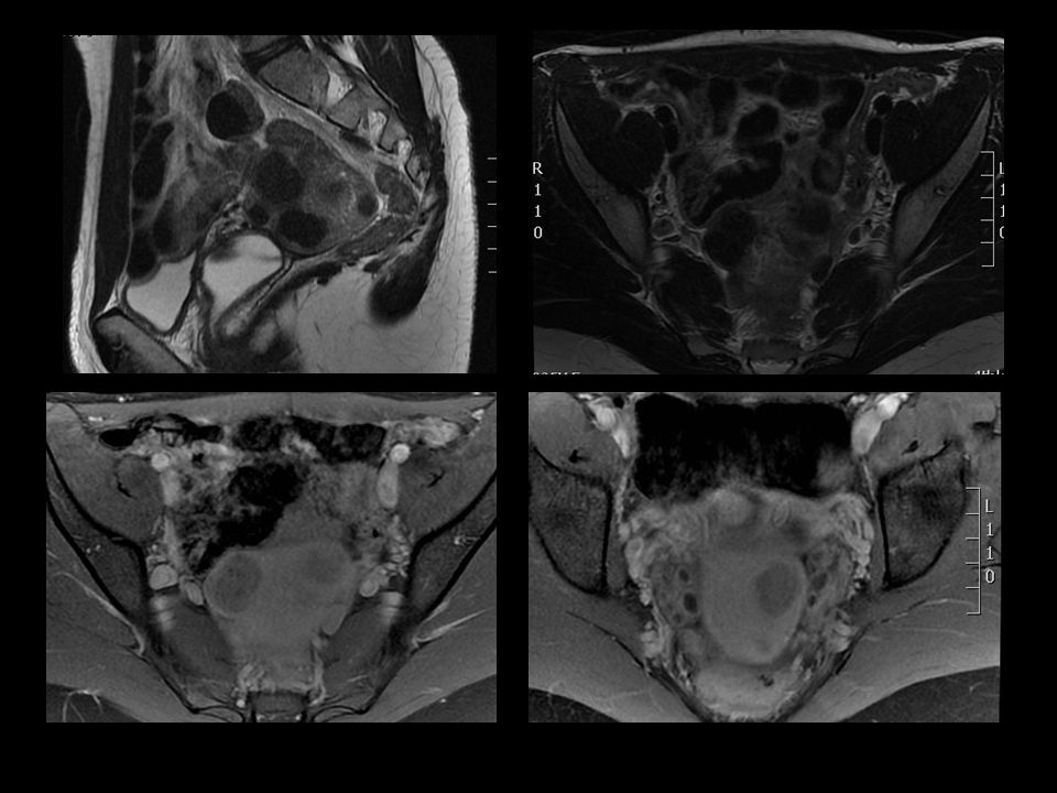
LEIOMIOMA SUBMUCOSO (SM).
Degenerated submucosal leiomyoma in a 46-year-old woman. Axial (a), coronal (b), oblique coronal (c), and sagittal (d) T2-weighted images show a degenerated leiomyoma (SM) that is located below the mucosal surface and protrudes into the endometrial cavity (arrows). Also note the nondegenerated submucosal leiomyoma (L) in b.
, coronal (b), oblique coronal (c), and sagittal (d) T2-weighted images show a degenerated leiomyoma (SM) that is located below the mucosal surface and protrudes into the endometrial cavity (arrows). Also note the nondegenerated submucosal leiomyoma (L) in b.")
9
LEIOMIOMA INTRACAVITÁRIO PEDUNCULADO (P)
LEIOMIOMA SUBSEROSO PEDUNCULADO (P) Pedunculated leiomyoma in a 40-year-old woman. Sagittal T2-weighted image shows a pedunculated intracavitary leiomyoma (P), which has prolapsed into the cervix on a long stalk (arrow). Deshmukh S P et al. Radiographics 2012;32:E251-E281
 Pedunculated leiomyoma in a 40-year-old woman. Sagittal T2-weighted image shows a pedunculated intracavitary leiomyoma (P), which has prolapsed into the cervix on a long stalk (arrow). Deshmukh S P et al. Radiographics 2012;32:E251-E281.")
10
LEIOMIOMA UTERINO INTRAMURAL Mais comum Assintomático Menorragia
Infertilidade (compressão cornoal / obstrui porção intersticial da tuba uterina)
")
12
LEIOMIOMA UTERINO Padrão histológico
Típico ou usual Celular Mitoticamente ativo Atípico Vascular Schwannóide Epitelióide Mixóide Lipoleiomioma PATOLOGISTA PROF. DRA Filomena Marino Carvalho

13
LEIOMIOMA CELULAR Musculatura lisa com pouco colágeno
Sem necrose celular Hiperintenso T2 Impregnação homogênea

14
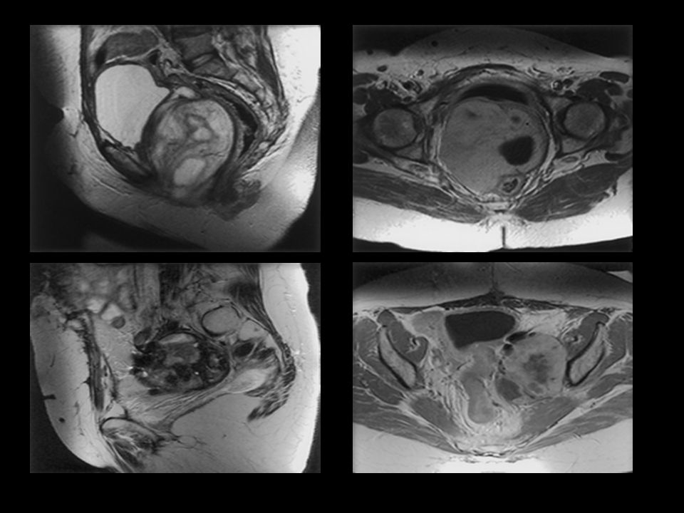
LEIOMIOMA CELULAR T2 T1 Gd
Pathologically proved cellular leiomyoma in a 35-year-old woman. (a) Axial T2-weighted image shows a large heterogeneous uterine mass with areas of increased signal intensity. (b) Axial contrast-enhanced fat-suppressed T1-weighted GRE image shows predominantly homogeneous enhancement of the mass. T T1 Gd
 Axial T2-weighted image shows a large heterogeneous uterine mass with areas of increased signal intensity. (b) Axial contrast-enhanced fat-suppressed T1-weighted GRE image shows predominantly homogeneous enhancement of the mass. T2 T1 Gd.")
15
Lipoleiomioma subseroso
T2 T1 T2 fat

16
Teratoma T2 T1 T1 fat pré T1 fat pós

17
LEIOMIOMA UTERINO RESSONÂNCIA MAGNÉTICA: LOCALIZAÇÃO TAMANHO NÚMERO
SUPRIMENTO VASCULAR COMPLICAÇÕES RM (CUSTO ELEVADO) X US < NÚMERO DE CIRURGIAS DESNECESSÁRIAS
 X US. < NÚMERO DE CIRURGIAS DESNECESSÁRIAS.")
18
LEIOMIOMA UTERINO TIPOS: DEGENERAÇÃO - INSUFICIÊNCIA VASCULAR
HIALINA MIXÓIDE CALCIFICADA CÍSTICA HEMORRÁGICA GORDUROSA DEGENERAÇÃO AGUDA (30 %): DOR/HEMORRAGIA
: DOR/HEMORRAGIA.")
19
LEIOMIOMAS DEGENERAÇÃO HIALINA
Leiomyomas with hyaline degeneration. Sagittal T2-weighted image shows two intramural leiomyomas with decreased signal intensity, a finding consistent with hyaline degeneration. (Courtesy of Evis Sala, MD, PhD, Memorial Sloan-Kettering Cancer Center, New York, NY.)
")
20
LEIOMIOMA DEGENERAÇÃO CÍSTICA (C)
Leiomyoma with cystic degeneration. Sagittal T2-weighted (a) and contrast-enhanced fat-suppressed T1-weighted GRE (b) images show an intracavitary leiomyoma that has undergone cystic degeneration. Note that the area of cystic degeneration (C) does not enhance after intravenous administration of gadolinium contrast material. The enlarged uterus causes mass effect on the urinary bladder (B). (Case courtesy of Evis Sala, MD, PhD, Memorial Sloan-Kettering Cancer Center, New York, NY.)
 and contrast-enhanced fat-suppressed T1-weighted GRE (b) images show an intracavitary leiomyoma that has undergone cystic degeneration. Note that the area of cystic degeneration (C) does not enhance after intravenous administration of gadolinium contrast material. The enlarged uterus causes mass effect on the urinary bladder (B). (Case courtesy of Evis Sala, MD, PhD, Memorial Sloan-Kettering Cancer Center, New York, NY.)")
21
LEIOMIOMA DEGENERAÇÃO MIXÓIDE

23
PRENHEZ ECTÓPICA NA CICATRIZ DE CESÁREA

24
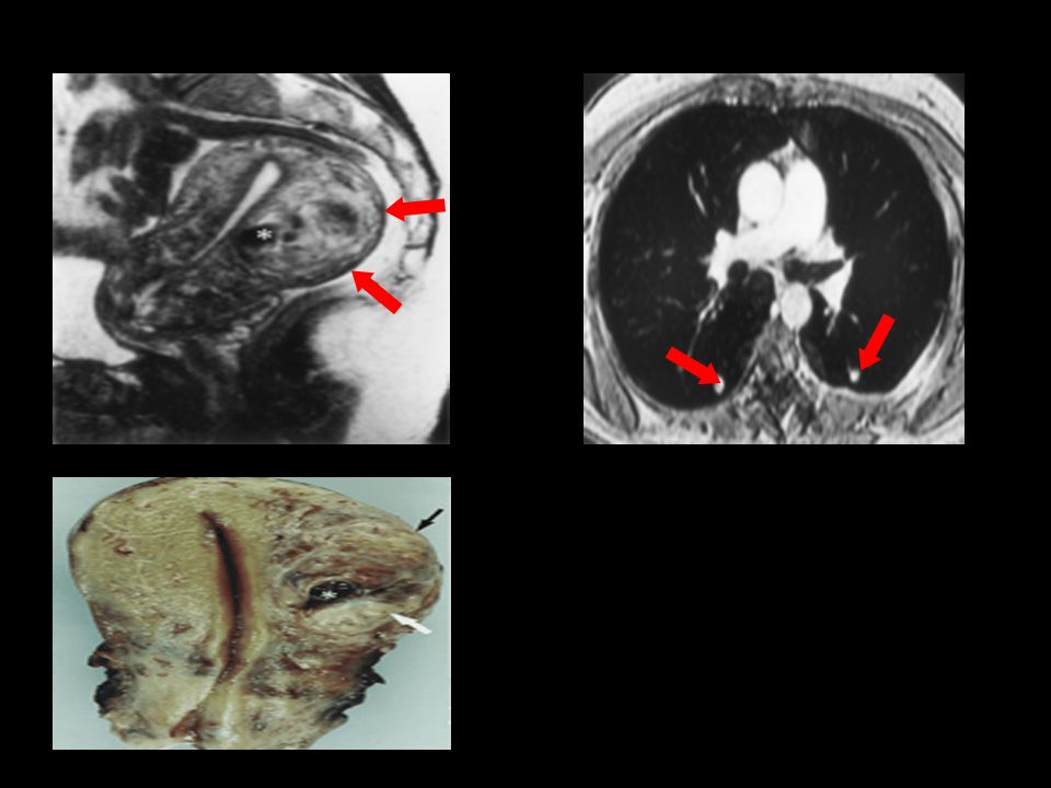
LEIOMIOMA DEGENERAÇÃO HEMORRÁGICA
Leiomyoma with red degeneration in a 32-year-old pregnant woman. (a, b) Axial (a) and sagittal (b) T2-weighted images show variable low signal intensity in a large leiomyoma (L) of the posterior lower uterine segment. Note the distortion and lengthening of the lower uterine segment and cervix (arrows in b) caused by the large leiomyoma. (c, d) Axial nonenhanced (c) and sagittal contrast-enhanced (d) fat-suppressed T1-weighted GRE images of the degenerated leiomyoma (L) show peripheral increased signal intensity (arrowheads) and a lack of contrast enhancement. T T1 FAT SAT
 Axial (a) and sagittal (b) T2-weighted images show variable low signal intensity in a large leiomyoma (L) of the posterior lower uterine segment. Note the distortion and lengthening of the lower uterine segment and cervix (arrows in b) caused by the large leiomyoma. (c, d) Axial nonenhanced (c) and sagittal contrast-enhanced (d) fat-suppressed T1-weighted GRE images of the degenerated leiomyoma (L) show peripheral increased signal intensity (arrowheads) and a lack of contrast enhancement. T2 T1 FAT SAT.")
25
LEIOMIOMA UTERINO PADRÃO DE CRESCIMENTO
LEIOMIOMATOSE INTRAVASCULAR LEIOMIOMATOSE PERITONEAL DISSEMINADA LEIOMIOMA TOSE METASTATIZANTE BENIGNA LEIOMIOMATOSE DISSECANTE (FORMA COTILEDONÓIDE) LEIOMIOMATOSE UTERINA LEIOMIOMA PARASITÁRIO (Suprimento arterial definido de uma estrutura pélvica ou abdominal adjacente).
 LEIOMIOMATOSE UTERINA. LEIOMIOMA PARASITÁRIO (Suprimento arterial definido de uma estrutura pélvica ou abdominal adjacente).")
26
—Benign LEIOMIOMATOSE METASTATIZANTE BENIGNA
—Benign metastasizing leiomyoma in 54-year-old woman who underwent hysterectomy 14 years earlier and was found to have incidental pulmonary nodules that had been stable for more than 10 years. Several nodules were resected soon after their initial incidental discovery for histologic diagnosis. IV contrast-enhanced axial CT image in lung windows shows numerous randomly distributed, smooth, rounded nodules (arrows) that are characteristic of benign metastasizing leiomyoma. Cohen D T et al. AJR 2007;188:
 that are characteristic of benign metastasizing leiomyoma. Cohen D T et al. AJR 2007;188:")
27
LEIOMIOMATOSE DISSECANTE (FORMA COTILEDONÓIDE)
LIGAMENTO LARGO Broad ligament leiomyoma in a 43-year-old woman. Coronal T2-weighted image shows a surgically proved leiomyoma (arrows) of the broad ligament. Although it is difficult to separate the leiomyoma from the uterus, it appears to be centered in the left broad ligament. Differential diagnostic considerations included an exophytic leiomyoma and a broad ligament leiomyoma.
 of the broad ligament. Although it is difficult to separate the leiomyoma from the uterus, it appears to be centered in the left broad ligament. Differential diagnostic considerations included an exophytic leiomyoma and a broad ligament leiomyoma.")
28
Leiomioma Adenomiose

29
LEIOMIOMAS (L) ADENOMIOSE (A)
Coexistence of adenomyosis and leiomyomas. Sagittal T2-weighted (a) and contrast-enhanced fat-suppressed T1-weighted GRE (b) images show a uterus containing adenomyosis (A in a) and multiple leiomyomas (L). At least one of the leiomyomas is pedunculated (P) with a short narrow stalk (arrow in a). (Case courtesy of Susan M. Ascher, MD, Georgetown University Hospital, Washington, DC.) Deshmukh S P et al. Radiographics 2012;32:E251-E281
 and contrast-enhanced fat-suppressed T1-weighted GRE (b) images show a uterus containing adenomyosis (A in a) and multiple leiomyomas (L). At least one of the leiomyomas is pedunculated (P) with a short narrow stalk (arrow in a). (Case courtesy of Susan M. Ascher, MD, Georgetown University Hospital, Washington, DC.) Deshmukh S P et al. Radiographics 2012;32:E251-E281.")
30
LEIOMIOMA X MASSA ANEXIAL LEIOMIOMA SUBSEROSO FIBROMA OVARIANO
Differentiation of a leiomyoma from an adnexal mass. Axial T2-weighted image shows a broad-based subserosal leiomyoma (L) with associated bridging vessels (arrow). The presence of bridging vessels allows the diagnosis of leiomyoma to be made, thereby allowing exclusion of an adnexal mass. (Courtesy of Evis Sala, MD, PhD, Memorial Sloan-Kettering Cancer Center, New York, NY.) LEIOMIOMA SUBSEROSO FIBROMA OVARIANO Deshmukh S P et al. Radiographics 2012;32:E251-E281
 with associated bridging vessels (arrow). The presence of bridging vessels allows the diagnosis of leiomyoma to be made, thereby allowing exclusion of an adnexal mass. (Courtesy of Evis Sala, MD, PhD, Memorial Sloan-Kettering Cancer Center, New York, NY.) LEIOMIOMA SUBSEROSO FIBROMA OVARIANO. Deshmukh S P et al. Radiographics 2012;32:E251-E281.")
31
CONTRAÇÃO UTERINA X LEIOMIOMA
Differentiation of a uterine contraction from a leiomyoma. Sagittal T2-weighted images show a transient uterine contraction (arrow in a). (Case courtesy of Kaori Togashi, MD, PhD, Kyoto University, Kyoto, Japan.)
. (Case courtesy of Kaori Togashi, MD, PhD, Kyoto University, Kyoto, Japan.)")
32
DIAGNÓSTICO DIFERENCIAL
METÁSTASE PARA ÚTERO Mama (lobular invasivo) e estômago (linite plástica): Infiltração difusa do corpo uterino Mama (carcinoma ductal invasivo) e melanoma: - Nodulações endometriais
 e estômago (linite plástica): Infiltração difusa do corpo uterino. Mama (carcinoma ductal invasivo) e melanoma: - Nodulações endometriais.")
33
LEIOMIOSSARCOMA UTERINO
1 – 2 % neoplasias malignas uterinas MULHERES 40 – 60 a ORIGEM: TECIDO CONECTIVO VASOS SANGUÍNEOS LEIOMIOMA PRÉ-EXISTENTE DE NOVO (MUSCULATURA UTERINA) TRANSFORMAÇÃO SARCOMATOSA NO LEIOMIOMA BENIGNO (0,1 – 0,8 %)
 TRANSFORMAÇÃO SARCOMATOSA NO LEIOMIOMA BENIGNO (0,1 – 0,8 %)")
34
LEIOMIOSSARCOMA UTERINO
Neoplasia de alto grau Tu agressivo Massa > 5,0 cm ZONA CENTRAL DE EXTENSA HEMORRAGIA / NECROSE FOCOS DE CALCIFICAÇÃO MARGEM IRREGULAR “ESPECIFICIDADE NÃO ESTABELECIDA DA RM”

35
LEIOMIOSSARCOMA UTERINO
Recidiva precoce local Crescimento rápido após menopausa Metástase hematogênica (pulmão, osso, cérebro) Linfonodos comprometidos é infrequente
 Linfonodos comprometidos é infrequente.")
36
LEIOMIOMA UTERINO CRITÉRIOS DE MALIGNIDADE
ATIPIA NECROSE CELULAR (FATOR PROGNÓSTICO) CONTAGEM MITÓTICA MIXÓIDE – CONTORNO INFILTRATIVO E ATIPIA EPITELIÓIDE – ATIPIA E NECROSE PATOLOGISTA PROF. DRA Filomena Marino Carvalho
 CONTAGEM MITÓTICA. MIXÓIDE – CONTORNO INFILTRATIVO E ATIPIA. EPITELIÓIDE – ATIPIA E NECROSE. PATOLOGISTA PROF. DRA Filomena Marino Carvalho.")
37
SARCOMA UTERINO NECROSE TUMORAL

38
LEIOMIOSSARCOMA UTERINO
RM: Inespecífica Grandes dimensões Sinal heterogêneo com áreas de hemorragia e/ou necrose Áreas de marcado realce tardio Invasão do miométrio/paramétrio Doença invasiva/metastática

41
LEIOMIOSSARCOMA UTERINO
Filomena Marino Carvalho LEIOMIOSSARCOMA UTERINO SUBTIPOS: CONVENCIONAL (CÉLULAS FUSIFORMES) MIXÓIDE EPITELIOIDE PATOLOGISTA PROF. DRA Filomena Marino Carvalho
 MIXÓIDE. EPITELIOIDE. PATOLOGISTA PROF. DRA Filomena Marino Carvalho.")
42
DIAGNÓSTICO DIFERENCAL
NEOPLASIA ESTROMAL ENDOMETRIAL Nódulo estromal Sarcoma estromal de baixo grau Sarcoma endometrial indiferenciado PATOLOGISTA PROF. DRA Filomena Marino Carvalho

43
DIAGNÓSTICO DIFERENCIAL
PECOMA – Tu de células epitelióides perivascular UTROSCT – “Tumores uterinos similares a tumores de cordões sexuais ovarianos” PATOLOGISTA PROF. DRA Filomena Marino Carvalho

44
DR. RONALDO RANGEL DR FABIO LEWIN

45
DR. RONALDO RANGEL DR. FABIO LEWIN

46
ADENOCARCINOMA SEROSO PAPILÍFERO DE ENDOMÉTRIO
DR. RONALDO RANGEL DR FABIO LEWIN

47
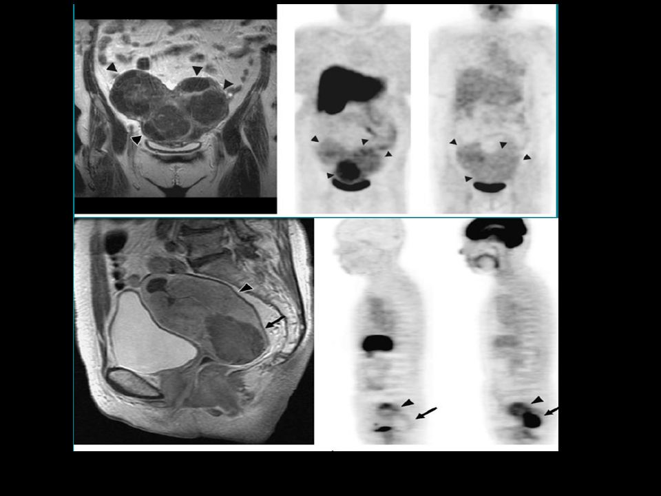
SARCOMA UTERINO Geralmente baixa/moderada captação pelo FDG
PET CT: LEIOMIOMA: Geralmente baixa/moderada captação pelo FDG Pode ter intensa captação pelo FDG (+ pcs pré-menopausa). LEIOMIOSSARCOMA: Moderada captação pelo FDG – difícil diferencial com mioma SARCOMA ENDOMETRIAL ESTROMAL: Intensa captação pelo FDG
. LEIOMIOSSARCOMA: Moderada captação pelo FDG – difícil diferencial com mioma. SARCOMA ENDOMETRIAL ESTROMAL: Intensa captação pelo FDG.")
49
LEIOMIOMA X SARCOMA UTERINO
OBRIGADO Douglas J. Racy

Apresentações semelhantes