Carregar apresentação
A apresentação está carregando. Por favor, espere

1
Métodos de diagnóstico: uma imagem vale mais do que mil palpites
Métodos gráficos Prof. Dr. Carlos Alberto Pastore

3
ELETROCARDIOLOGIA

4
Monitorização Ambulatorial do Eletrocardiograma
A partir de 1961 quando NORMAN J. HOLTER introduziu na prática clínica os equipamentos para monitoração ambulatorial do ECG. Sua utilização tem sido crescente.

5
MONITORIZAÇÃO AMBULATORIAL DO ELETROCARDIOGRAMA
HOLTER LOOPER

6
TESTE ERGOMÉTRICO

7
MAPEAMENTO ELETROCARDIOGRÁFICO DE SUPERFÍCIE
BODY SURFACE POTENTIAL MAPPING ELETROCARDIOLOGIA / INCOR-HCFMUSP

8
CENTRAL DE TRANSMISSÃO

9
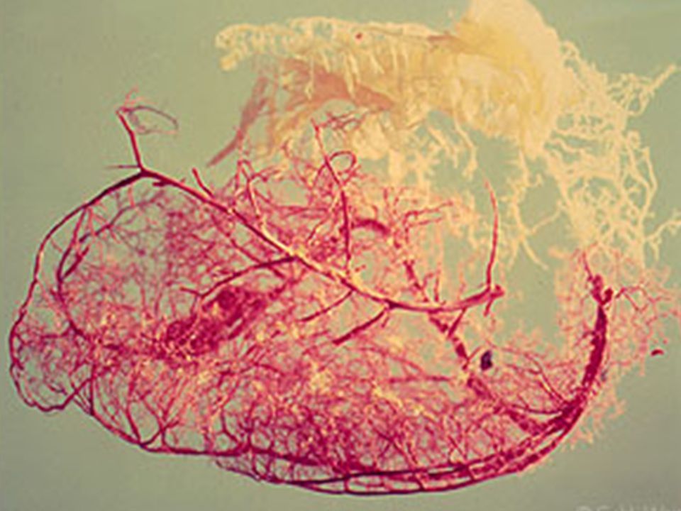
ARTERIOESCLEROSE ATEROSCLEROSE

11
Andersen [22]; with permission.)
Figure 3-7. Macroscopic view of a vulnerable plaque. A, The yellow, soft, atheromatous gruel is separated from the vascular lumen by only a thin, but intact, fibrous cap. The vascular lumen is distended by white radiographic contrast medium injected post mortem. B, This cross-sectional specimen was just a few millimeters distal to the one shown in A. The thin fibrous cap is ruptured, a big cap fragment and some of the soft atheromatous gruel are missing (owing to downstream embolization), and a mural thrombus has evolved where the thrombogenic atheromatous gruel has been exposed. White contrast medium has penetrated the soft gruel through the ruptured cap. (Adapted from Falk and Andersen [22]; with permission.) ©Copyright Science Press Internet Services
![Andersen [22]; with permission.)](http://slideplayer.com.br/slide/3676907/12/images/11/Andersen+%5B22%5D%3B+with+permission.%29.jpg "Figure 3-7. Macroscopic view of a vulnerable plaque. A, The yellow, soft, atheromatous gruel is separated from the vascular lumen by only a thin, but. intact, fibrous cap. The vascular lumen is distended by white radiographic. contrast medium injected post mortem. B, This cross-sectional specimen was just. a few millimeters distal to the one shown in A. The thin fibrous cap is. ruptured, a big cap fragment and some of the soft atheromatous gruel are missing. (owing to downstream embolization), and a mural thrombus has evolved where the. thrombogenic atheromatous gruel has been exposed. White contrast medium has. penetrated the soft gruel through the ruptured cap. (Adapted from Falk and. Andersen [22]; with permission.) ©Copyright Science Press Internet Services.")
12
TC Multislice Anatomia Coronária Normal

13
Angioplastia coronária

14
MEDICINA NUCLEAR

15
CINTILOGRAFIA MIOCÁRDICA DE PERFUSÃO

16
SPECT

17
L.M.F., 65 anos, Masculino, Angor Atípico, TE com ST.
REPOUSO PÓS ESTRESSE FEVE= 52% FEVE= 46%

18
PET: PERFUSÃO E METABOLISMO
UCLA 1998

19
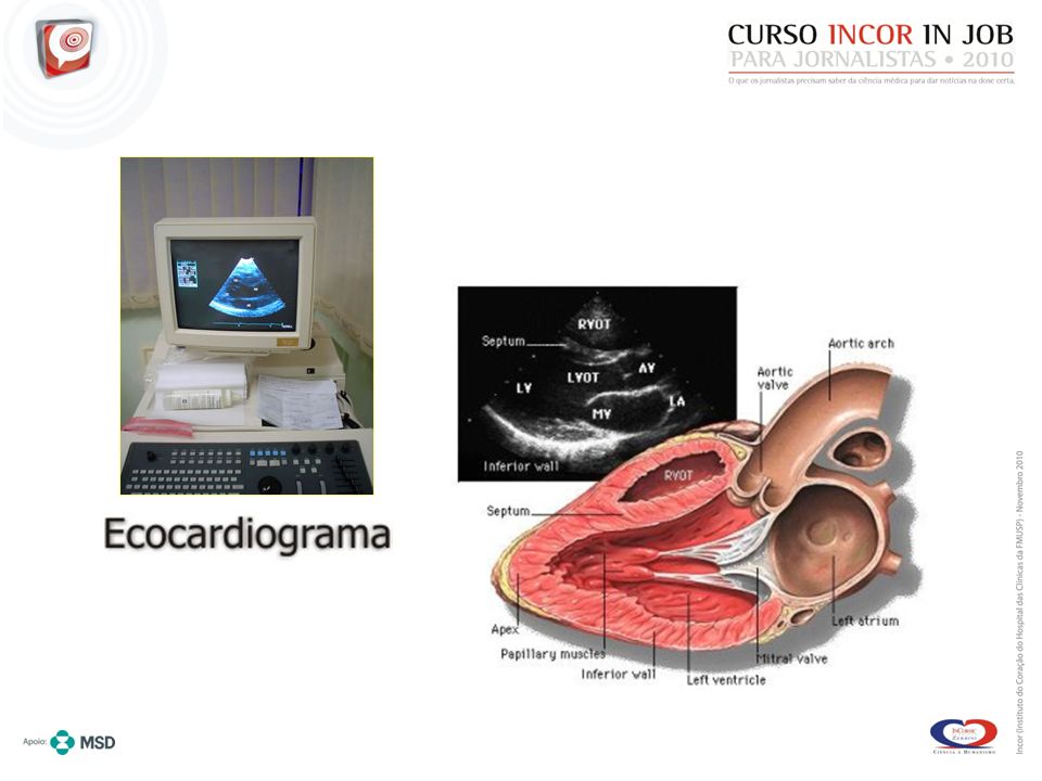
ECOCARDIOGRAMA Ultrasom

20
Ultra-som Renal

22
Repouso Adenosina Dobutamina
RFM = 6,2 RFM = 6,8

23
Insuficiência Cardíaca QRS largo DISSINCRONIA RESSINCRONIZAÇÃO

24
Ressincronização Cardíaca

27
Ressincronização Cardíaca MARCAPASSO BIVENTRICULAR

28
Estimulação Biventricular Técnica Transvenosa
And in this other na combination of atrio-biventricular pacing with defibrillator. PA Perfil

29
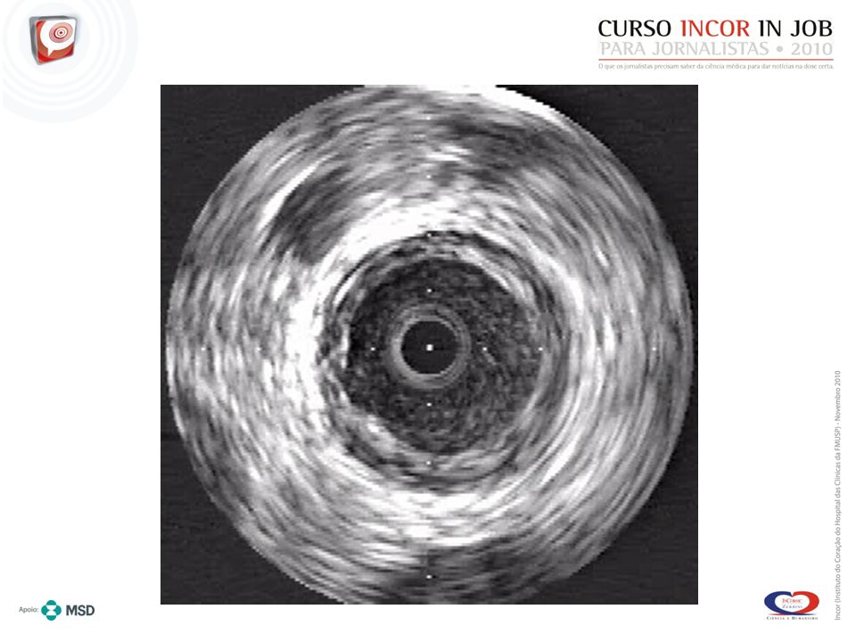
ULTRASOM – INTRAVASCULAR
PLACA DURA x PLACA MOLE

30
Ultra-Som Intravascular

32
Placa Aterosclerótica com Ruptura
Figure (see Color Plate) Additional examples of ruptured aortic atherosclerotic plaques with exposure of their lipid cores (LC) to blood elements and subsequent thrombosis. A, The ruptured ends of the fibrous cap (FC) (arrows) are shown to be considerably thinner than the rest of the collagenous intima over the plaque. Masson trichrome, original magnification, x80. B, The extruded atheromatous debris is transformed into a sizable luminal thrombus (original magnification, x200).Although the relation between atherosclerosis and thrombosis has been recognized for more than 100 years, its importance has been downplayed. In recent years, an important link between atherosclerosis and thrombosis has been attributed to lipoprotein(a) [Lp(a)]. This lipoprotein is composed of a low-density lipoprotein particle linked by a disulfide bridge to a unique apoprotein, apo(a). Apo(a) is strikingly homologous (75% to 90%) to human plasminogen. Because of this strong homology, Lp(a) binds to fibrin, whereupon it competes with both plasminogen and tissue-type plasminogen activator (t-PA) for fibrin binding, thereby reducing the significant enhancement in t-PA`s catalytic efficiency that fibrin binding facilitates [40]. Lp(a) has been demonstrated by immunohistochemistry in atheromatous lesions. By downregulating plasmin generation, Lp(a) leads to impaired activation of transforming growth factor (TGF)-B and thus may contribute to smooth muscle cell proliferation. Lp(a) may also contribute to atherogenesis by participating in the control of angiogenesis. This latter action of Lp(a) may convert a stable to an unstable plaque. Furthermore, Lp(a) may be involved in the recruitment of monocytes to the arterial wall [41]. Finally, it has been shown that patients with high levels of Lp(a) have impairment of endothelial-dependent vasodilation of epicardial arteries [42]. A–adventitia; M–media; PH–plaque hemorrhage; T–thrombus (with cholesterol clefts). ©Copyright Science Press Internet Services
 Additional examples of ruptured aortic. atherosclerotic plaques with exposure of their lipid cores (LC) to blood. elements and subsequent thrombosis. A, The ruptured ends of the fibrous cap (FC) (arrows) are shown to be considerably thinner than the rest of the collagenous. intima over the plaque. Masson trichrome, original magnification, x80. B, The. extruded atheromatous debris is transformed into a sizable luminal thrombus. (original magnification, x200).Although the relation between atherosclerosis and. thrombosis has been recognized for more than 100 years, its importance has been. downplayed. In recent years, an important link between atherosclerosis and. thrombosis has been attributed to lipoprotein(a) [Lp(a)]. This lipoprotein is. composed of a low-density lipoprotein particle linked by a disulfide bridge to a. unique apoprotein, apo(a). Apo(a) is strikingly homologous (75% to 90%) to human. plasminogen. Because of this strong homology, Lp(a) binds to fibrin, whereupon. it competes with both plasminogen and tissue-type plasminogen activator (t-PA) for fibrin binding, thereby reducing the significant enhancement in t-PA`s. catalytic efficiency that fibrin binding facilitates [40]. Lp(a) has been. demonstrated by immunohistochemistry in atheromatous lesions. By downregulating. plasmin generation, Lp(a) leads to impaired activation of transforming growth. factor (TGF)-B and thus may contribute to smooth muscle cell proliferation. Lp(a) may also contribute to atherogenesis by participating in the control of. angiogenesis. This latter action of Lp(a) may convert a stable to an unstable. plaque. Furthermore, Lp(a) may be involved in the recruitment of monocytes to. the arterial wall [41]. Finally, it has been shown that patients with high. levels of Lp(a) have impairment of endothelial-dependent vasodilation of. epicardial arteries [42]. A–adventitia; M–media; PH–plaque. hemorrhage; T–thrombus (with cholesterol clefts). ©Copyright Science Press Internet Services.")
33
Classificação de Placa Vulnerável
Normal PV c/Erosão PV Propensa a Ruptura PV Propensa a Erosão PV c/Ruptura/Cicatrizando PV com nódulo calcificado PV criticamente estenótica PV com hemorragia intra-placa

34
SITES RECOMENDADOS

35
PUBLICAÇÕES RECOMENDADAS
Eletrocardiologia Atual Editora/Ano de publicação: Editora Atheneu, 2009

Apresentações semelhantes

>")


